
News|Articles|December 18, 2025
Test Detects Single Circulating Tumor Cells Using Infrared Imaging
Author(s)Alexandra Gerlach, Content Producer
Fact checked by: Nicole Canfora Lupo
Listen
0:00 / 0:00
Key Takeaways
- A novel blood test using infrared technology detects individual cancer cells, potentially transforming lung cancer detection and monitoring.
- The method identifies circulating tumor cells (CTCs) by their unique chemical fingerprints, offering a less invasive and more affordable alternative to current tests.
A UK research team has pioneered a blood test using infrared technology to detect lung cancer cells, promising earlier diagnoses and personalized treatments.
Advertisement
A team of researchers from the UK developed a blood test capable of detecting individual cancer cells, potentially shifting how clinicians detect and monitor disease in lung cancer. The approach is unique—utilizing infrared technology to identify circulating tumor cells (CTCs).1
“Our team was able to detect a single lung cancer cell in a patient’s blood by combining advanced infrared scanning technology with computer analysis, focusing on the unique chemical fingerprint of cancer cells,” said Josep Sulé-Suso, PhD, associate specialist in oncology at UHNM and lead author of the study.1
“This approach has the potential to help patients receive earlier diagnoses, personalized treatments, and fewer invasive procedures, and it could eventually be applied to many types of cancer beyond lung cancer."1
Lung Cancer in the United States
Lung cancer is the leading cause of cancer death in the US, with fatalities surpassing those of breast, prostate, and colorectal cancers combined. Treatment advances have greatly improved outcomes for patients—with 5-year survival rates exceeding 60% if the disease is localized.2 However, lung cancer screening uptake is low around the country, highlighting a critical care gap and opportunities to create diagnostic approaches to increase screening availability.
CTC Detection in Lung Cancer
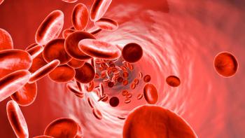
Detection of CTCs is a growing diagnostic and monitoring method across malignancies, showing promising capabilities in detecting cancer using less invasive testing. CTCs are cancer cells that can break away from tumors and travel in the bloodstream. As a method for testing, they offer vital clues about disease progression and treatment response.
Despite the early success of CTC detection, available testing methods are complex, expensive, and time-consuming. In some cases, these tests can miss cancer cells, as they often change their characteristics in the bloodstream.
To address these limitations, Sulé-Suso and his team developed a method to detect CTCs in blood samples using infrared light. When shining a highly concentrated beam of infrared light onto the samples, they found that different chemicals absorb infrared light in different ways. CTCs showed a distinct absorption pattern, or "chemical fingerprint."
The finding has the potential to revolutionize access to and affordability of effective lung cancer screening—providing a simpler and more affordable technique that is easier to adopt into everyday clinical practice.
“Professionally, it offers the chance to translate fundamental spectroscopy into meaningful medical impact,” said Sulé-Suso. “Personally, it resonates strongly: cancer has affected my own family and claimed the lives of friends, and helping to expand the tools available to fight this disease gives me a powerful sense of purpose.”
“The possibility that our work could one day influence clinical practice, reduce diagnostic delays, and improve patient outcomes is a constant driving force behind this research.”
Building the Next Generation of Lung Cancer Detection Tools
The study presents a first proof-of-concept for Fourier transform infrared (FT-IR) microspectroscopy, combined with a random forest classifier. The team analyzed cytospun blood samples for the detection of a single CTC in a lung cancer patient. Notably, the method employs standard glass coverslips routinely used in pathology workflows, allowing for straightforward integration with conventional histopathological techniques, including staining and immunohistochemistry.3
Using FT-IR spectral data derived from in vitro–cultured lung cancer cells as the training set, the approach achieved accurate CTC identification based on biochemical signatures within the fingerprint region (1800–1350 cm-1). These findings establish FT-IR microspectroscopy as a novel, label-free strategy for CTC detection in liquid biopsies, with the potential to expand the diagnostic landscape in oncology.
“Contributing to research with the potential to transform early cancer detection is both a professional privilege and a deeply personal motivation,” said Paul Roach, PhD, an expert in biomaterials and interface science at Loughborough University. “The possibility that our work could one day influence clinical practice, reduce diagnostic delays, and improve patient outcomes is a constant driving force behind this research.”1
REFERENCES
1. Researchers develop groundbreaking blood test for lung cancer. Loughborough University. December 16, 2025. Accessed December 18, 2025. https://www.lboro.ac.uk/media-centre/press-releases/2025/december/groundbreaking-blood-test-lung-cancer/
2. Gerlach A. Age-based lung cancer screening could dramatically improve early detection. November 28, 2025. Accessed December 18, 2025. https://www.pharmacytimes.com/view/age-based-lung-cancer-screening-could-dramatically-improve-early-detection
3. Dowling L, Evans C, Roach P, et al. Fourier transform infrared microspectroscopy as a liquid biopsy tool to detect single circulating tumour cells in the blood of a lung cancer patient. Applied Spectroscopy. Published online October 15, 2025. doi:10.1177/00037028251390565
Advertisement
Related Content
Advertisement


Latest CME


Advertisement
Advertisement
Trending on Pharmacy Times
1
Cyclosporiasis Cases Reach 5100: Investigators Zero in on Lettuce, Possible Taco Bell Link
2
FDA Approves At-Home Starting Dose of Lecanemab for Treatment of Alzheimer Disease
3
Psychedelics in Cluster Headaches Are Breaking the Pain Cycle
4
Medicare and Weight-Loss Medications: A Pharmacist's FAQ on the GLP-1 Bridge Program
5