
|Articles|November 29, 2022
Study Highlights Changes to the Brain Caused by Migraines
Author(s)PT Staff
Research into how perivascular spaces contribute to migraine could help better understand the complexities of how these headaches occur.
Advertisement
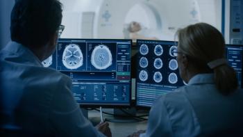
New research indicates that migraine headaches can cause changes to the brain that have never been observed before. The study, presented at the Radiological Society of North America annual meeting, identified enlarged perivascular spaces—spaces filled with fluid that surround the brain’s blood vessels—in the brains of patients with migraine.
Perivascular spaces are most commonly found in the basal ganglia and white matter of the cerebrum, as well as along the optic tract.
“In people with chronic migraine and episodic migraine without aura, there are significant changes in the perivascular spaces of a brain region called the centrum semiovale,” study co-author Wilson Xu, an MD candidate at Keck School of Medicine of the University of Southern California, said in a press release. “These changes have never been reported before.”
Migraines can cause nausea, weakness, and sensitivity to light. More than 37 million US residents are affected by migraine, according to the American Migraine Foundation.
Perivascular spaces are influenced by factors such as abnormalities at the blood-brain barrier and inflammation. Enlarged perivascular spaces can be a sign of underlying small vessel disease, according to the study authors.
“Perivascular spaces are part of a fluid clearance system in the brain,” Xu said. “Studying how they contribute to migraine could help us better understand the complexities of how migraines occur.”
The researchers sought to evaluate the link between migraine and enlarged perivascular spaces using an ultra-high field 7T MRI to compare structural microvascular changes in different types of migraines.
“To our knowledge, this is first study using ultra-high resolution MRI to study microvascular changes in the brain due to migraine, particularly in perivascular spaces,” Xu said in the release. “Because 7T MRI is able to create images of the brain with much higher resolution and better quality than other MRI types, it can be used to demonstrate much smaller changes that happen in brain tissue after a migraine.”
The study enrolled 10 patients with chronic migraine, 10 patients with episodic migraine without aura, and 5 age-matched healthy patients in the control group. All patients were between 25 and 60 years of age. Exclusion criteria were overt cognitive impairment, brain tumor, prior intracranial surgery, MRI contraindications, and claustrophobia.
Enlarged perivascular spaces were measured in the centrum semiovale and basal ganglia regions of the brain. White matter hyperintensities were analyzed using the Fazekas scale and cerebral microbleeds were measured with the microbleed anatomical rating scale.
Additionally, the investigators gathered clinical data on disease duration and severity, symptoms at time of scan, presence of aura, and side of headache.
The data showed that the number of enlarged perivascular spaces in the centrum semiovale was significantly greater in patients with migraine than in the control group. Further, enlarged perivascular space quantity in the centrum semiovale was associated with deep white matter hyperintensity severity in patients with migraine.
“We studied chronic migraine and episodic migraine without aura and found that, for both types of migraine, perivascular spaces were bigger in the centrum semiovale,” Xu said. “Although we didn’t find any significant changes in the severity of white matter lesions in patients with and without migraine, these white matter lesions were significantly linked to the presence of enlarged perivascular spaces. This suggests that changes in perivascular spaces could lead to future development of more white matter lesions.”
The significant differences in the perivascular spaces in patients with migraine vs the healthy control group may suggest glymphatic disruption within the brain; however, it is unknown whether these changes influence the development of migraine or result from migraine, according to the study authors. The added that future research with larger case populations and longitudinal follow-up is needed to better establish the link between structural changes and migraine development and type.
“The results of our study could help inspire future, larger-scale studies to continue investigating how changes in the brain’s microscopic vessels and blood supply contribute to different migraine types,” Xu said. “Eventually, this could help us develop new, personalized ways to diagnose and treat migraine.”
Reference
Ultra-high-res MRI Reveals Migraine Brain Changes. Radiological Society of North America. News release. November 23, 2022.
Advertisement
Related Content
Advertisement


Latest CME


Advertisement
Advertisement
Trending on Pharmacy Times
1
FDA Panel Unanimously Backs Moderna’s Breakthrough mRNA Flu Vaccine Amid Political Turbulence
2
Sonrotoclax Plus Zanubrutinib Achieves Undetectable MRD Rates Above 90% in Frontline CLL/SLL
3
ADA 2026: Lipid Management and Cardiovascular Prevention Updates for People With Diabetes
4
McKesson ideaShare 2026: How AI and Automation Are Reshaping the Dispensing Experience
5