
|Articles|July 18, 2016
Novel Imaging Technique Can Quickly Distinguish Tumor Grade
Author(s)Laurie Toich, Assistant Editor
RSI-MRI can distinguish prostate tumor grade quickly and accurately.
Advertisement
Findings from a recent study suggest that using restriction spectrum imaging (RSI) as a biomarker can enhance the differentiation of prostate tumors with magnetic resonance imaging (MRI).
"Noninvasive imaging is used to detect disease, but RSI-MRI takes it a step further," said study senior author David S. Karow, MD, PhD. "We can predict the grade of a tumor sometimes without a biopsy of the prostate tissue. This is taking all that's good about multi-parametric MRI and making it better."
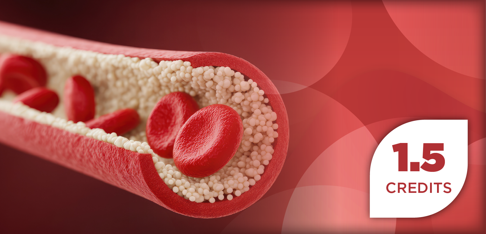
Researchers found that adding RSI to a pelvic MRI only added 2.5 to 5 minutes of scanning time, making it fast, accurate. The procedure showed a decreased risk compared with traditional MRI, which requires the patient to be injected with dye, according to the study published in Clinical Cancer Research.
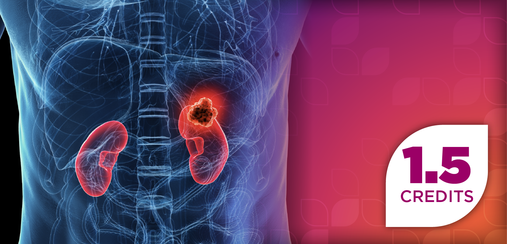
RSI-MRI corrects magnetic field distortion and focuses on water diffusion in tumor cells that have a high nuclear volume fraction, according to the study. Through this process, researchers are able to more accurately find the tumor’s location and decipher the tumor’s grade.
The study noted that RSI-MRI can guide treatment and biopsy to the target region with the highest-grade tumor. Over 1000 patients received RSI-MRI imaging, and patients had MR-fused ultrasound guided prostate biopsy.
"Previously, we relied completely on systematic, but random, biopsies of the prostate to diagnose cancer, which has been the standard practice in our field for years. Now, we use RSI-MRI to precisely target specific areas of concern and enhance the accuracy of our diagnosis," said researcher J. Kellogg Parsons, MD, MHS, UC. "Greater accuracy means improved care tailored to each individual patient. With RSI-MRI, we are better able to identify which cancers are more aggressive and require immediate treatment, and which ones are slow growing and can be safely observed as part of a program called active surveillance."
For this study, researchers included 10 patients and evaluated more than 2700 data points. Researchers believe that the next steps would be to introduce this imaging to other hospitals to see if it can be used alone.
According to the study, RSI-MRI as a stand-alone non-contrast screening tool could take 15 minutes for a contrast-enhanced exam that would typically last up to 1 hour.
"What our evidence shows so far is the imaging benefit is coming from RSI-MRI," concluded study senior author Dr Karow. "I think this technique could become standard of care and mainstream for the vast majority of men who are at risk for prostate cancer. Full contrast MRI is expensive and risky for most men. This is the kind of exam that could be done on a routine clinical basis."
Newsletter
Stay informed on drug updates, treatment guidelines, and pharmacy practice trends—subscribe to Pharmacy Times for weekly clinical insights.
Advertisement
Related Content
Advertisement


Latest CME


Advertisement
Advertisement
Trending on Pharmacy Times
1
Polycystic Ovary Syndrome Undergoes Name Change: Polyendocrine Metabolic Ovarian Syndrome
2
Ketamine’s Expanding Role in Chronic Pain and Opioid-Sparing Therapy
3
Navigating GLP-1 Access: A Pharmacy Technician’s Role
4
Pharmacy Care Under Pressure: Pairing Standard Practice with Always-On Surveillance for Safer, More Efficient Care
5