
News|Articles|June 2, 2023
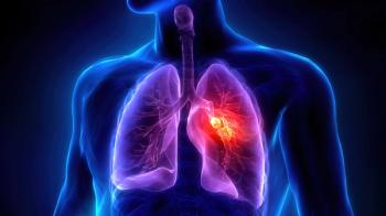
COVID-19 Infection Could Be Detected in Lungs, Heart, Causing Inflammatory Damage
Author(s)Ashley Gallagher, Editor
Investigators found thinner ventricular walls, disorganized and ruptured myocardial fiber, mild inflammatory infiltration, and mild epicardia or interstitial fibrosis in the hearts of mice infected with COVID-19.
Advertisement
Viral RNA may be detected in the lungs and hearts of mice infected with the Beta variant of COVID-19, according to the results of a non-transgenic mouse model study published in Antiviral Research. Investigators also found that
During a pathological analysis, researchers found that there were thinner ventricular walls, disorganized and ruptured myocardial fiber, mild inflammatory infiltration, and mild epicardia or interstitial fibrosis in the hearts of the infected mice. The investigators used 3 mutations, K417N, E484K and N501Y, which helped to facilitate the recognition of the Beta variant of COVID-19 within the mice. It also helped to promote viral infection in the wild-type mice.
A COVID-19 infection caused apoptosis, a reduction of mitochondrial integrity and quality, as well as cessation of beating within the human pluripotent stem cell-derived cardiomyocyte-like cells, according to the investigators. They sought to determine the mechanism of the myocardial injury caused by a COVID-19 infection.
Investigators used transcriptome sequencing of the human pluripotent stem cell-derived cardiomyocyte-like cells at different time points during the infection.
The analysis revealed a large induction of inflammatory cytokines and chemokines, upregulations of major histocompatibility complex class 1 molecules, activation of apoptosis signaling, and cell cycle arresting. Investigators believe that this could be the cause of inflammation, immune cell infiltration, and cell death. In addition, the inflammation could be caused by the infection releasing cytokines and chemokines in the human pluripotent stem cell-derived cardiomyocyte-like cells.
Based on the findings, investigators called for a biological regulation for COVID-19-associated cardiomyopathy due to the infection reducing the contractility of cardiomyocytes, cell growth, and apoptosis. The investigators determined that there were several genes associated with cell cycle regulation that decreased in the infection of cardiomyocytes, including CDK1, CDK6, and CCNE2. They said that this could have an effect on cell cycle arrest.
The study also found that captopril, a hypotensive drug targeting angiotensin-converting enzyme (ACE), could help to alleviate the inflammatory response, as well as apoptosis in the cardiomyocytes by inactivating the tumor necrosis factor (TNF) signaling pathways, including TNF-NFKB, TNF-p38 MAPK and TNF-JNK. The investigators said that these findings suggest that captopril may be beneficial for reducing COVID-19-associated cardiomyopathy.
Captopril increases ACE2 levels as well as anti-inflammatory activity, with the investigators citing that ACE inhibitors do not increase an individual’s susceptibility to COVID-19 severity or morality.
Investigators noted that typically, upregulated expression of ACE2 can lead to increased susceptibility to COVID-19, but captopril did not have the same increased risk. They added that the mechanisms around captopril have yet to be determined, but one possible mechanism could be due to ACE2 shedding.
They said that these findings may offer a preliminary explanation into the mechanism of pathological cardiac injury associated with COVID-19 and provide a new perspective for discovery and development on antiviral therapeutics.
Reference
Huang X, Fan W, Sun J, Yang J, et al. SARS-CoV-2 induces cardiomyocyte apoptosis and inflammation but can be ameliorated by ACE inhibitor Captopril. Antiviral Res. 2023;215:105636. doi:10.1016/j.antiviral.2023.105636
Advertisement
Related Content
Advertisement


Latest CME


Advertisement
Advertisement
Trending on Pharmacy Times
1
FDA Approves Enlicitide, the First Oral PCSK9 Inhibitor for High Cholesterol
2
The Weekly Dose: Cyclosporiasis Outbreak Grows, AI-Powered Refills Pilot, and New FDA Approvals in Alzheimer Disease
3
GLP-1s and Oncology: The Drug Interaction Clinicians Aren't Talking About
4
Improving Detection and Management of ATTR-CM Through Pharmacy Care
5