|Articles|October 22, 2021
Pharmacy Practice in Focus: Oncology
- October 2021
- Volume 3
- Issue 5
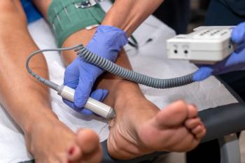
Antineoplastic Extravasation Prevention, Management

Extravasation is defined as the inadvertent leakage of a vesicant from the vein into the surrounding tissue.
Advertisement
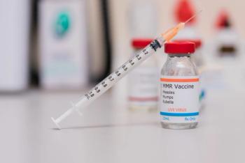
Although infiltration can be defined as the inadvertent leakage of an irritant, extravasation is defined as the inadvertent leakage of a vesicant from the vein into surrounding tissue. During cancer treatment, extravasation is the accidental leakage of chemotherapy from the vein into surrounding tissue.1,2 Rates are not clearly defined and no centralized database of chemotherapy extravasation events exists, but some literature reports overall incidence as ranging from 0.1% to 6.5%.3,4
The damage potential from antineoplastic therapy extravasation ranges from mild toxicity, such as local pain and erythema, to severe toxicity including tissue necrosis. Because of the potential for severe tissue injury, it is imperative that preventive measures are taken and extravasation is identified and managed quickly.1,5
Ensuring team members are educated about what causes extravasation, opportunities to prevent it, and how to respond when chemotherapy leakage occurs is critically important. There are a few concepts that pharmacists working in oncology need to know about extravasation involving commonly used chemotherapies.
Classification of Agents
Chemotherapy drugs are grouped according to their potential to cause tissue damage if extravasation occurs; agents may be classified as irritants or vesicants. Depending on the treatment being utilized, other categories such as nonvesicant, nonirritant, or irritant with vesicant-like properties may be used as well.1,3,5
Classifications of antineoplastic drugs are not absolute. Chemotherapy agents may have characteristics of irritants and vesicants.6 Additionally, there are inconsistencies among antineoplastic agent classifications in the literature.1,5
If extravasation of an irritant occurs, there is potential for burning, pain, and erythema. This is typically a temporary, local reaction without tissue necrosis.5,7,8 Vesicants have potential for burning, pain, and erythema, along with more severe, progressive damage including blister formation and tissue necrosis.
Vesicants can be further subclassified into DNA-binding and non–DNA-binding agents. DNA-binding vesicants remain bound to the DNA of dead cells and can be internalized by adjacent, healthy cells. This allows the agent to remain in the tissue, continuing to cause damage. The damage potential of DNA-binding vesicants is generally more severe, progressive, and permanent.
Non–DNA-binding vesicants are metabolized in the tissue and damage potential is generally mild to moderate, localized, and improves with time.5,7,8 Examples of DNA-binding vesicants are anthracyclines, whereas examples of non–DNA-binding vesicants are vinca alkaloids.1 Table 1 shows the European Society for Medical Oncology and European Oncology Nursing Society Clinical Practice Guideline classification of chemotherapy drugs.1
Risk Factors
Identifying and acknowledging extravasation risk factors is essential to reducing risk. Risk factors may be classified as patient-related, procedure-related, or drug therapy-related, as noted in Table 2.1,2,5,8
Prevention
Implementation of prevention measures is key to minimizing risk of extravasation and potential consequences, with education on these measures remaining crucial for the medical staff and the patient. Medical staff should be trained in risk identification, appropriate vascular access, institute protocols, and extravasation prevention, identification, management, and documentation. Additionally, patients should be educated on extravasation risk and the need to promptly report any signs or symptoms of its occurrence.1,5,7
The vascular access site options include central or peripheral access, with central access being generally preferred for chemotherapy agents, especially those with more severe damage potential such as anthracyclines. A flexible cannula should be used, and large veins of the forearm are preferred when selecting a cannulation site for peripheral access.1
Identification and Management
The clinical presentation of an extravasation may present as a wide range of symptoms that are nonspecific, such as tingling, burning, discomfort, pain, swelling, erythema, and visible accumulation of fluid. Additionally, there may be a lack of blood return, resistance on the syringe plunger, or an interruption in the free flow of the drug infusion.1,5
If an extravasation of an antineoplastic agent is identified, general management principles are as follows1:
- Stop the infusion
- Disconnect tubing while leaving the cannula in place
- Without applying pressure, carefully aspirate as much drug as possible using a syringe
- Remove the cannula
- Assess the site and outline border of extravasation area with a pen
- Notify physician
- Initiate substance-specific measures
- Thermal compresses
- Antidotes, if applicable
- Elevate limb
- Documentation
- Follow-up
Dry, thermal compresses should be applied in 20-minute intervals 4 times a day for 1 to 2 days.5 There are some inconsistencies among thermal compress recommendations pending the resource utilized.
However, most agents will require cold compresses for extravasation management. For extravasation of vinca alkaloids, warm compresses are universally recommended.1,5 Substance-specific antidotes should also be used as appropriate (Table 3).1,3,5
If dexrazoxane is being administered for anthracycline extravasation management, it must be started within 6 hours after extravasation. Additionally, cold compresses should be removed 15 minutes before and during dexrazoxane administration to maximize antidote delivery to the tissue.1
The documentation of extravasation incidents should include the patient’s name, date and time of extravasation, name of extravasated drug and diluent used, signs and symptoms, IV access utilized, extravasation area, and management steps.
Best practice recommendations for documentation also include incorporating photographic documentation, because this can be helpful during follow-up. Patients should be instructed to monitor the area and report changes. Routine follow-up is recommended with more frequent monitoring within the first week post extravasation, followed by weekly review until symptoms are resolved.1
Surgical Intervention
Surgical debridement may be necessary if patients experience unresolved tissue necrosis or pain lasting more than 10 days, full-thickness skin necrosis, and/or chronic ulcer. For severe tissue damage, it is recommended that surgical intervention be managed by a plastic surgeon. Necrotic tissue will be excised, followed by skin grafting and reconstruction if necessary.1
Antineoplastic therapy extravasation is a serious event that has the potential for severe complications. Employing strategies and preventive interventions will help ensure extravasation incidents remain rare and that staff are prepared to respond promptly and appropriately.
Christina Billias, PharmD, is a clinical pharmacy specialist at Roswell Park Comprehensive Cancer Center in Buffalo, New York.
Megan Menon, PharmD, BCOP, is a medication safety officer at Roswell Park Comprehensive Cancer Center in Buffalo, New York.
REFERENCES
1. Perez Fidalgo JA, Garcia Fabregat L, Cervantes A, Margulies A, Vidall C, Roila F. Management of chemotherapy extravasation: ESMO-EONS clinical practice guidelines. Ann Oncol. 2012;23(suppl 7):167-173. doi:10.1093/annonc/mds294
2. Le A, Patel S. Extravasation of noncytotoxic drugs: a review of the literature. Ann Pharmacother. 2014;48(7):870-886. doi:10.1177/1060028014527820
3. Jackson-Rose J, Del Monte J, Groman A, et al. Chemotherapy extravasation: establishing a national benchmark for incidence among cancer centers. Clin J Oncol Nurs. 2017;21(4):438-445. doi:10.1188/17.CJON.438-445
4. Ener RA, Meglathery SB, Styler M. Extravasation of systemic hemato-oncological therapies. Ann Oncol. 2004;15(6):858-862. doi:10.1093/annonc/mdh214
5. Boulanger J, Ducharme A, Dufour A, Fortier S, Almanric K;
6. Howell G, Oliai C, Schiller G. Liposomal cytarabine-daunorubicin (CPX-351) extravasation: case report and literature review. Anticancer Res. 2018;38(12):6927-6930. doi:10.21873/anticanres.13070
7. Kreidieh FY, Moukadem HA, El Saghir NS. Overview, prevention and management of chemotherapy extravasation. World J Clin Oncol. 2016;7(1):87-97. doi:10.5306/wjco.v7.i1.87
8. Schulmeister L. Extravasation management: clinical update. Semin Oncol Nurs. 2011;27(1):82-90. doi:10.1016/j.soncn.2010.11.010
Articles in this issue
over 4 years ago
Improving Adherence to Oral Oncolyticsover 4 years ago
Updates in Prostate Cancer Treatmentover 4 years ago
CAR T Cell–Associated CRS, Neurotoxicityover 4 years ago
Overview of PARP Inhibitors in the Treatment of Ovarian CancerAdvertisement
Related Content
Advertisement


Latest CME


Advertisement
Advertisement
Trending on Pharmacy Times
1
Medicare's New GLP-1 Bridge Program: What Pharmacists Need to Know Before Dispensing
2
Isatuximab Wins FDA Approval, Becoming Second Subcutaneous Anti-CD38 for Multiple Myeloma
3
FDA Expands Wilate Approval for Routine Prophylaxis in Children Younger Than 6 With von Willebrand Disease
4
Single-Shot Respiratory Vaccine Could Transform Pharmacist Counseling and Immunization Rates
5