
|Articles|September 1, 2006
- Volume 0 0
Antiplatelet Agents in the Prevention of Atherothrombotic Events
Author(s)Quinn Wells, MD, PharmD
Advertisement
For full disclosure information, send an e-mail request to: [email protected].
Behavioral Objectives
After completing this continuing education article, the pharmacist should be able to:
- Discuss the epidemiology as well as the personal and societal cost of cardiovascular disease (CVD).
- Describe the pathophysiology of CVD with particular attention to the role of platelets.
- Characterize the pharmacology of the commonly used oral platelet active agents used for the prevention of cardiovascular events.
- Understand and describe the current evidence and recommendations regarding the use of oral antiplatelet prophylaxis.
Cardiovascular disease (CVD), including stroke, is the leading cause of death and disability in the United States and is responsible for a great deal of morbidity, mortality, lost productivity, and health care costs. The pathophysiology of CVD is complex and remains to be completely elucidated. Central, however, to the most devastating manifestations of the disease is the process of atherothrombosis, or acute thrombus formation secondary to the rupture or erosion of an underlying atherosclerotic lesion.
Thrombus formation, in turn, is critically dependent on platelets. Much effort has been expended to understand the role of platelets in atherothrombosis and to develop agents that can disrupt platelet function in the hope of interrupting the sequence of events that lead to occlusive thrombi.
A number of antiplatelet agents have been investigated for the primary and secondary prevention of atherothrombotic events. Whereas there are some data for dipyridamole, the most convincing evidence is available for low-dose aspirin and clopidogrel. Both agents have been shown to reduce the incidence of vascular events in a variety of patient populations, and each also has been shown to increase the risk of serious bleeding. For these reasons, it is important to identify patients in whom the benefit of primary or secondary prophylaxis with these agents is sufficient to offset the risks of therapy.
Cost and Burden of CVD to Patients and Society
More than 70 million Americans currently live with CVD, and more than 910,000 die each year from it. The most common forms of CVD are heart disease and stroke. They are the first and third leading causes of death in the United States, respectively. Combined, they account for nearly 40% of deaths annually. 1-3 Furthermore, coronary artery disease is the leading cause of premature, permanent disability in the US workforce. Stroke alone accounts for about 1 million cases of disability in this country. Including direct health care costs and lost productivity, the cost of heart disease and stroke in the United States is projected to exceed $400 billion in 2006.2,3
Pathophysiology of CVD: A Focus on Platelets
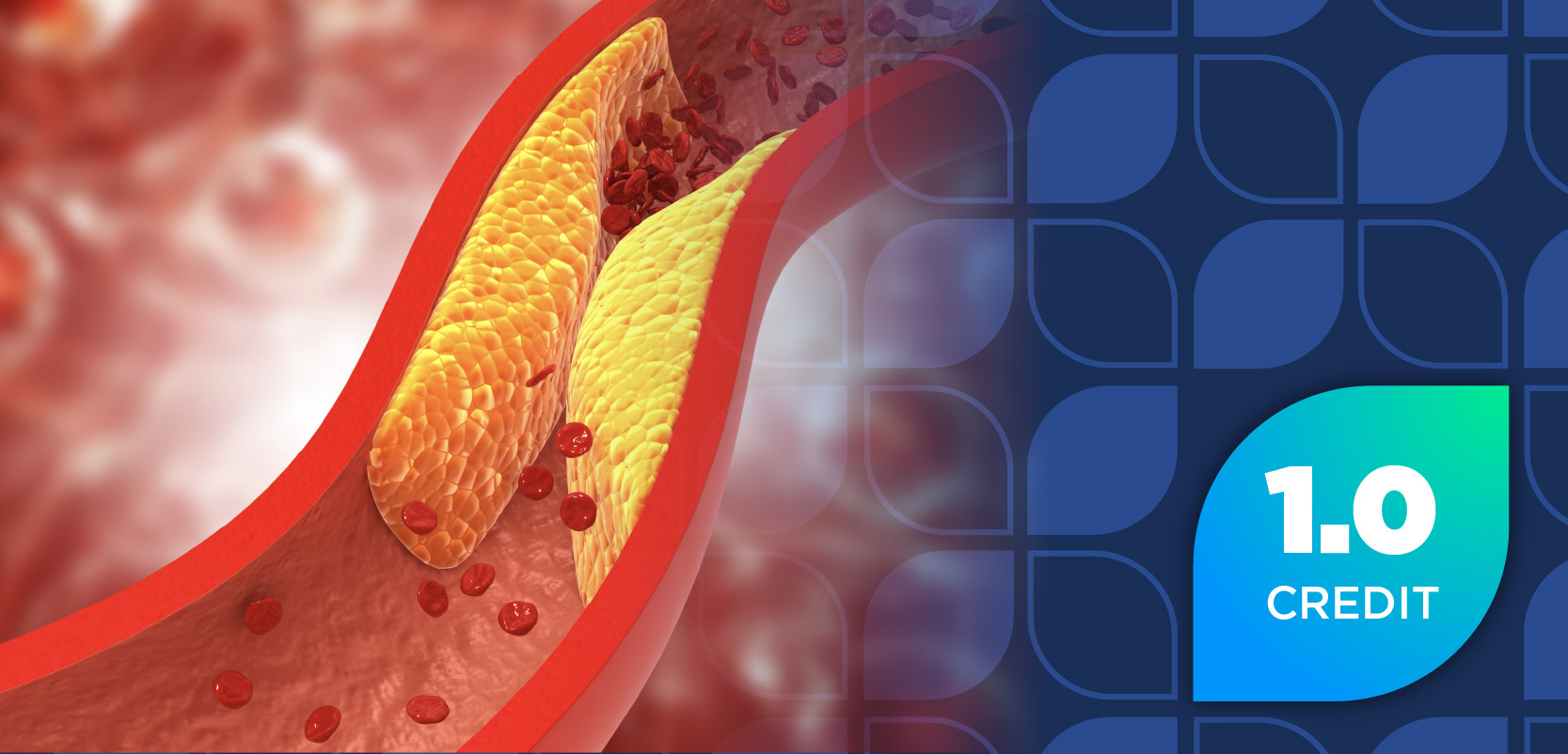
Although all forms of CVD cause some degree of morbidity and/or mortality, the most deadly manifestation of CVD is arterial thrombosis. This is an acute complication that develops on the chronic lesion of atherosclerosis.4 The formation of the atherosclerotic plaque is a complicated process that occurs over the course of years. The rupture or erosion of this plaque leads to the rapid formation of a clot that can compromise blood flow to downstream structures.
The understanding of this process has given rise to the term atherothrombosis to describe both the chronic and the acute processes of arterial disease.4,5 Arterial thrombosis causes vascular compromise and leads to the clinical manifestations of acute coronary syndromes (ie, unstable angina and myocardial infarction [MI]) and ischemic stroke.
A great deal of effort has been put into understanding the pathogenesis of the atherosclerotic lesion, as well as the process of acute thrombus formation. This research has revealed that acute arterial thrombosis, a crucial event in the most devastating of CVD, is intimately intertwined with platelet activity.4
The Atherosclerotic Lesion
The underlying lesion of atherothrombosis is the atherosclerotic plaque. The development of the atherosclerotic plaque is a complex event involving the endothelium, smooth muscle cells, macrophages, T-lymphocytes, and platelets.6
Risk factors for the development of atherosclerosis include elevated lowdensity lipoprotein (LDL) cholesterol, cigarette smoking, hypertension, diabetes, male gender, increasing age, hyperhomocysteinemia, obesity, and physical inactivity.5,6
Of these risk factors, only elevated LDL cholesterol seems able to drive the development of atherosclerosis alone. The other factors appear to augment and accelerate the process being driven by atherogenic lipoproteins.5
Autopsies of young, asymptomatic individuals also have demonstrated that, as the number of cardiovascular (CV) risk factors increases, so does the severity of coronary and aortic atherosclerosis.7 Conversely, exercise, elevated high-density lipoprotein cholesterol, and moderate alcohol consumption appear to protect against the development of atherosclerosis.5,8,9
Atherosclerotic lesions begin to develop when lipoproteins from the plasma extravasate through leaky, dysfunctional endothelium (the innermost layer of arteries) into the subendothelial space. Atherogenic lipoproteins are retained and modified, primarily by oxidation, to become proinflammatory, atherogenic, cytotoxic, and chemotactic.5,10,11 In response to the accumulation of proatherosclerotic material, the endothelium becomes activated and expresses a number of adhesion molecules that facilitate the recruitment of immune cells from the plasma and thus initiate the inflammatory component of atherogenesis.5,12
Among the cells recruited to the site are monocytes. After migration into the lesion, they differentiate into macrophages and begin phagocytosing the oxidized LDL. Interestingly, macrophages ingest this altered LDL using an alternate receptor (the so-called scavenger receptor), which is not downregulated by high intracellular LDL concentrations as is the native LDL receptor. Thus, they continue to ingest oxidized LDL until they become engorged and are known as foam cells. With a continued supply of oxidized LDL, macrophages continue to phagocytose oxidized LDL until they die. The accumulation of dead, lipid-laded macrophages contributes to the development of the soft, lipid-rich core characteristic of atherosclerotic lesions.5 Early, asymptomatic foam cell lesions are the characteristic "fatty streaks" of early atherosclerosis.
In response to the injury imposed by the above processes, a fibroproliferatory phase begins. The smooth muscle cells of the vessel primarily mediate this phase in an effort to control and repair damage. The smooth muscle cells migrate around the lipid-rich core and secrete a collagen-rich matrix. This action forms a fibrous cap separating the core from the flow of blood. The persistent stimulus of the lesion leads to an exuberant response and growth of the lesion. As disease progresses, calcification is common in atherosclerotic lesions.13,14
Plaques that are stable may enlarge and compromise the lumen of the affected vessel. This situation gives rise to the syndromes of peripheral arterial disease (eg, claudication) and stable angina.
A more ominous course occurs when the fibrous cap to the plaque either erodes or, more commonly, ruptures, exposing the thrombogenic lipid core to the circulation. This rupture sets in motion a series of events that leads to thrombus formation and acute arterial occlusion either at the plaque site or downstream by emboli. This process gives rise to stroke and the acute coronary syndromes of unstable angina and MI.
Platelet Physiology
Platelets are anucleated, discoid blood cells that derive from megakaryocytes in the bone marrow. The life span of a typical platelet is, on average, ~10 days in the circulation. Under physiologic conditions, platelets in the circulation are able to distinguish between normal intact endothelium and injured endothelium.
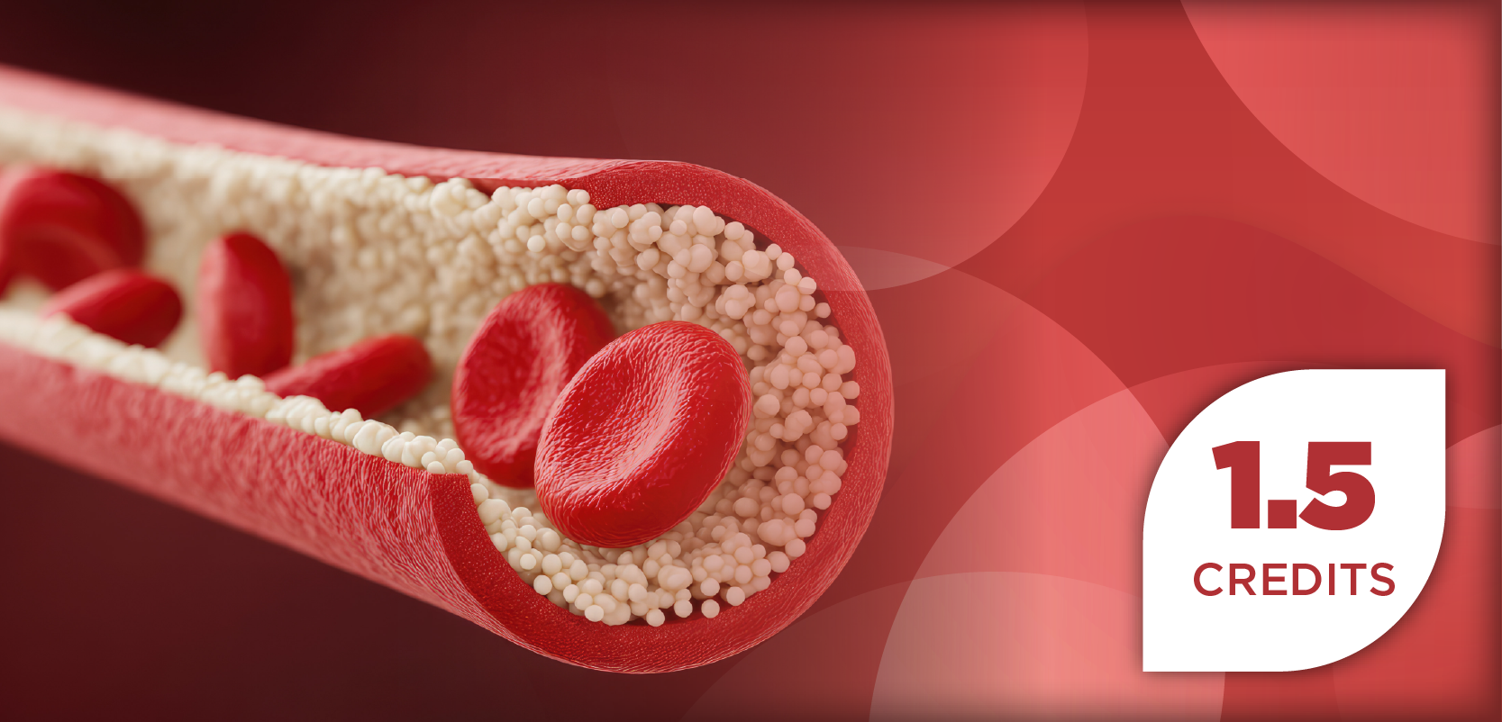
When a lesion is identified, platelets adhere to the site, become activated, and recruit other platelets to the location. This process also promotes the accumulation of fibrinogen, the precursor of fibrin, at the developing clot. Under normal conditions, this process facilitates hemostasis after traumatic disruption of the vasculature. The same process, however, can lead to arterial occlusion and downstream tissue damage. Thus, a delicate balance between rapid hemostasis and blood fluidity exists in the body.
A key player in this process is the endothelium.4 Endothelial cells control vascular tone and help to regulate hemostasis by secreting a number of factors that both inhibit and promote platelet function and blood clotting.15,16 The primary stimulus for thrombus formation is the loss of endothelium and the resultant exposure of underlying extracellular matrix components. Following this event, platelets undergo sequential and coordinated steps of adhesion, activation, and aggregation.4
Understanding the role of antiplatelet therapy requires an understanding of the molecular events involved in normal platelet function. There are a dizzying array of substrates, agonists, ligands, receptors, and signaling pathways that orchestrate the complex physiology of platelets.
Platelet Adhesion
When the endothelium of a vessel is disrupted, a number of subendothelial matrix constituents are exposed that support platelet adhesion. Principally involved are collagens and von Willebrand factor, although fibronectin, laminins, and other substances participate.4,17 Platelets have membrane-bound receptors for each of these adhesive proteins that allow interactions between the platelet and the subendothelial matrix. Furthermore, a single ligand may interact with multiple surface receptors, and, conversely, a single receptor may interact with multiple ligands.17 The foregoing interactions mediate the first stage of hemostasis?the physical closure of defects in vessel walls by the adherence of platelets.
Platelet Activation
The initial interaction between platelet receptors and subendothelial constituents facilitates adhesion. These interactions, however, also initiate signaling pathways that lead to platelet activation.
Platelet activation is a complex process marked by a number of events. First, the cytoskeleton of platelets reorganizes in such a way that the typical discoid shape is lost, and they become irregularly shaped and develop cytoplasmic extensions. Second, activated platelets begin to secrete the contents of preformed granules into the cytosol (eg, calcium ions) and the extracellular space (eg, von Willebrand factor and adenosine diphosphate [ADP]).
Other events include the liberation of arachidonic acid from the cell membrane by phospholipase A2. Once released, arachidonic acid is acted on by cyclooxygenase-1 (COX-1) to form intermediate prostaglandins, which in turn are acted on by thromboxane-A synthase to form thromboxane A2 (TXA2). These substances act locally to reinforce platelet activation and accentuate aggregation.4,15,17 Both TXA2 and ADP activate surface receptors that, when stimulated, act locally to further platelet recruitment and activation. Thus, they are key amplifiers of the initial steps of hemostasis.
Another key event in platelet activation is the conversion of a surface glycoprotein, GP IIb/IIa, from an inactive form to an active form. As described below, this glycoprotein plays a key role in platelet aggregation and clot formation.
A final characteristic of activated platelets is the surface expression of anionic phospholipids that serve as binding and activation sites for components of the coagulation cascade. Thus, the activated platelet serves to link platelet plug formation to the coagulation cascade.
Platelet Aggregation and Clot Formation
The various stimuli that may mediate adherence and stimulate activation all converge on the common pathway of aggregation mediated by the nowactive form of GP IIb/IIa. Activated GP IIb/IIa binds to a number of adhesive proteins, most notably fibrinogen, but also to von Willebrand factor and others. GP IIb/IIa from adjacent platelets may interact with the same fibrinogen molecule. In this fashion, platelets begin to aggregate and form substrate onto which other platelets are recruited and activated. Within minutes, this process leads to the formation of a platelet plug at the site of injury.
As discussed previously, activated platelets display particular phospholipids on their surfaces that serve as binding sites for factors in the coagulation cascade. Once activated, the coagulation system results in the production of the enzyme thrombin, which converts fibrinogen to fibrin. The deposition of fibrin results in a fibrin-platelet meshwork and transforms the initial platelet plug into a stable clot.4,17
Aspirin
Mechanism of Action and Pharmacokinetics
Aspirin, or acetylsalicylic acid, is perhaps the best known of currently available antiplatelet agents. Aspirin has been used for many years as an analgesic, anti-inflammatory agent, and antipyretic. In the 1940s, the hemorrhagic effects of aspirin were noted. In the 1960s, it was discovered that these effects were related to platelets, and 20 years later the importance of prostaglandins was elucidated.18
Aspirin is rapidly absorbed in the stomach and upper small intestines by diffusion. With regular aspirin, peak plasma concentrations are achieved in 30 to 40 minutes with a bioavailability of 40% to 50%. Enteric-coated formulations have a considerably lower bioavailability, and peak concentrations may not occur for 3 to 4 hours.19
Aspirin exerts its antiplatelet activity by inactivating COX, the enzyme that catalyzes the first committed step in prostaglandin synthesis. This step involves the conversion of membranederived arachidonic acid to prostaglandin H2. This intermediate, in turn, is converted by multiple tissue-specific enzymes to at least 5 biologically active compounds, including TXA2 and prostacycline.20 Platelets are particularly rich in thromboxane synthase, the enzyme responsible for the production of TXA2.
By interrupting this pathway, aspirin prevents the thromboxane-mediated platelet activation outlined above. Molecularly, aspirin first binds to an arginine residue located at position 120 in the enzyme. This location is the common docking site for all nonsteroidal anti-inflammatory drugs (NSAIDs). Aspirin then irreversibly acetylates a serine residue (located at position 529 in COX-1 and position 516 in COX-2), which prevents arachidonic acid from gaining access to the catalytic site.21 Platelets are initially exposed to aspirin in the portal circulation and are therefore exposed to higher concentrations than those present in the peripheral circulation.
Aspirin has a plasma half-life of 15 to 20 minutes; however, the antiplatelet effects persist for the lifetime of affected platelets. Such is the case because of the irreversibility of the COX inactivation and the anuclear nature of platelets. This second fact means that platelets are incapable of synthesizing new enzymes to replace those inactivated by aspirin. Because of the disconnect between the pharmacokinetics and pharmacodynamics of aspirin, oncedaily regimens of low-dose aspirin generally are sufficient to provide a substantial antiplatelet effect.20-22
Efficacy
The efficacy of aspirin has been documented in a broad spectrum of patient populations. These groups range from patients free of CVD to those presenting with acute MI or stroke. Whereas aspirin has consistently been shown to reduce the incidence of both fatal and nonfatal vascular events (death from a vascular event, nonfatal stroke, or MI), the magnitude of these effects varies according to the clinical setting.
It is useful to review the data in terms of primary versus secondary prevention. Important trials investigating the use of aspirin as primary prevention are shown in Table 1.
In patients with very low baseline risk of CVD, the benefits of aspirin are offset by the risk of aspirin-induced bleeding. As the risk of a CV event increases, however, the risk-benefit ratio of therapy becomes more favorable. Likewise, as the baseline risk of adverse events increases (eg, in older patients or in those taking other NSAIDs), a higher risk of CV events is required to justify aspirin therapy. Thus, given the known risk of aspirin therapy (discussed below), the decision to implement aspirin for primary prophylaxis of CVD requires the assessment of the 5-to 10-year risk of experiencing a CV event.22 Correspondingly, the clinician also must make an assessment of the risk of drug therapy in each patient.
In addition, a number of trials have demonstrated the effectiveness of aspirin in the setting of acute CV events (eg, MI, unstable angina, and ischemic stroke) and in the secondary prevention of CV events in patients with established disease. In general, aspirin reduces the risk of a serious vascular event (nonfatal MI, nonfatal stroke, or death from any vascular event) by ~25% in patients with established disease. This estimate is a composite of a 34% reduction in the rate of nonfatal MI, a 25% decrease in the rate of nonfatal stroke, and an ~17% decrease in the death rate.20
The most comprehensive analysis of the available data has been performed by the Antithrombotic Trialists' Collaboration.23 This extensive meta-analysis demonstrated a clear benefit from aspirin use in a number of high-risk population groups. Data from this analysis are shown in Figure 1. These data translate into the prevention of ~10 to 40 vascular events per 1000 high-risk patients treated per year. Figure 2 shows the benefit of therapy in groups with varying baseline risk.
Safety/Side Effects
The safety of aspirin in various populations has been evaluated by a number of authors. The major side effect of aspirin is bleeding. The most common site of serious bleeding (ie, requiring hospitalization or transfusion) is the gastrointestinal (GI) tract. Intracranial hemorrhage is a less common, but deadly, complication of aspirin therapy. Estimates from available data suggest that treatment with low-dose aspirin in high-risk patients approximately doubles the risk of major extracranial bleeding. For nonelderly patients, this risk translates into 1 to 2 major hemorrhagic events per 1000 patients treated per year. There is an estimated increase of 1 to 2 intracranial hemorrhages per 10,000 patients treated per year.20 Although not detected by the Antithrombotic Trialists' Collaboration meta-analysis, there are data that support the notion that the risk of GI toxicity is dose-dependent. This risk seems to be slightly less with enteric-coated formulations.
Furthermore, the risk of a major bleeding event is higher in the elderly, with patients >70 years of age at the greatest risk.22 For example, in one study in which the average age of patients receiving aspirin was 73.9 years, the average age of patients who experienced a complication was 79.7 years.4 The risk of GI hemorrhage is increased by a history of GI disturbance (GI pain, complicated or uncomplicated ulcer).20 Some gastroenterologists have advocated the use of proton pump inhibitors in high-risk patients taking low-dose aspirin.24 Clearly, patients at low risk for experiencing vascular events have a poorer risk-benefit ratio for aspirin therapy.
Dosing Issues
The GI toxicity of aspirin seems to be dose-dependent. Furthermore, there is evidence that suggests that higher doses of aspirin are actually less effective at preventing atherothrombosis. This observation is in line with the fact that very low doses of aspirin are able to selectively inhibit platelet COX-1, and thus thromboxane synthesis, whereas higher doses of aspirin begin to inhibit endothelial COX-1 and COX-2. These enzymes are responsible for the production of endothelial prostacycline, which has potent antithrombotic effects. For these reasons, it is recommended that the clinician use the lowest dose that has been shown to be effective for the condition22 (Table 2).
Clopidogrel
Mechanism of Action and Pharmacokinetics
Clopidogrel, along with ticlopidine, is a member of the thienopyridine class of antiplatelet drugs.18,22 Ticlopidine has fallen out of favor because it is associated with neutropenia (1%-2.4%) and thrombotic thrombocytopenia purpura (1 in 3000). Ticlopidine also requires twice-daily dosing. Following administration, clopidogrel is rapidly absorbed from the GI tract and is metabolized by hepatic cytochrome P-450 enzymes. After single oral doses of up to 200 mg, or repeated daily doses of up to 100 mg, unchanged clopidogrel is not detectable in the circulation. Clopidogrel has no in vitro antiplatelet activity, and it is thought that hepatic conversion to an active metabolite is necessary for pharmacologic activity.
Thienopyridines exert their antiplatelet effects by irreversibly inhibiting the P2Y12 receptor on the platelet membrane. The endogenous ligand for this receptor is ADP, and when activated it mediates ADP-dependent platelet aggregation. Like aspirin, clopidogrel causes changes that last for the life span of the platelet. Also as with aspirin, small daily doses are sufficient to cause considerable platelet dysfunction. Following discontinuation, normal platelet physiology returns in ~1 week, owing to the production of new platelets to replace those altered by clopidogrel.18,25,26
Efficacy
The efficacy of clopidogrel was established in the Clopidogrel Versus Aspirin in Patients at Risk of Ischaemic Events (CAPRIE) study.27 This trial enrolled >19,000 patients with recent cerebrovascular accident, MI, or symptomatic peripheral artery disease who were followed over a period of up to 3 years (average follow-up 1.91 years). Clopidogrel 75 mg daily was compared with aspirin 325 mg daily, with a primary end point of a composite of stroke, MI, or vascular death.
In the CAPRIE study, there was a small, but statistically significant, reduction in the primary end point (5.32% vs 5.83%; P = .043). The most pronounced benefits were seen in patients with peripheral artery disease. In this trial, the incidence of bleeding was similar in aspirin and clopidogrel patients (approximately 9%), although GI bleeding was more common in the aspirin group.
Following these results, the Management of Atherothrombosis with Clopidogrel in High-Risk Patients with Recent Transient Ischaemic Attack or Ischaemic Stroke (MATCH) study investigated whether the addition of aspirin to clopidogrel provided increased benefit.28 The trial randomized nearly 7600 patients with recent transient ischemic attack or ischemic stroke and at least one additional risk factor to receive clopidogrel plus aspirin (75 mg/day) or placebo. The patients were followed for 18 months, with a primary end point of a composite of ischemic stroke, MI, vascular death, or rehospitalization for acute ischemia or worsening of peripheral artery disease.
This trial showed a small, nonsignificant benefit from dual therapy (15.7% vs 16.7%; P = .24). There was, however, a doubling in the risk of major bleeding associated with combination therapy (2.6% vs 1.3%; P < .001), although there was no change in mortality. As a consequence of these results, dual therapy in these patients has been reconsidered.
The Clopidogrel in Unstable Angina to Prevent Recurrent Ischemic Events (CURE) trial investigated the effects of clopidogrel in addition to aspirin in patients with non-ST-segment elevation acute coronary syndromes.29 This study enrolled >12,500 patients who presented within 24 hours of symptoms. There was a 20% relative risk reduction in the composite end point of nonfatal MI, death, or stroke (11.4% vs 9.3%; P < .001). The risk of major bleeding was higher in the dual-treatment group (3.7% vs 2.7%; P = .001), although the difference in life-threatening bleeding was not significant (2.2% vs 1.8%; P = .13). The role of clopidogrel in ST-segment elevation MI also has been evaluated (the CLARITY study).30 All patients received fibrinolytics, aspirin, and heparin. Patients were randomized to receive either placebo or clopidogrel in addition to standard care.
This trial showed a 36% reduction (21.7% vs 15%) in the composite end point of need for revascularization, recurrent MI, or death. There was no difference in the incidence of major bleeding.
A special niche for clopidogrel has been in patients who have experienced an acute coronary syndrome with or without percutaneous coronary intervention (PCI) with stent placement. In this setting, data have shown that combination therapy with aspirin and clopidogrel for several months post stent placement provides benefit.
Safety/Side Effects
As with aspirin, the major side effect of clopidogrel is bleeding. In randomized trials, the risk of major bleeding with clopidogrel was similar to that seen with aspirin therapy. When it is used in combination with aspirin, however, the result is a significant increase in the risk of serious bleeding. Therefore, this approach is used only in select groups of patients (eg, patients who have undergone PCI), and, even in these groups, combination therapy is used for a limited time.
In the CAPRIE trial, the discontinuation rate for clopidogrel was similar to that with aspirin (12%). Clopidogrel was associated with higher rates of rash and diarrhea, whereas aspirin was associated with higher rates of GI discomfort. Clopidogrel was not associated with excess neutropenia or thrombocytopenia.22,27 Although thrombotic thrombocytopenia purpura has been associated with clopidogrel, it is rare and typically occurs in the first 2 weeks of therapy.31
Dipyridamole + Aspirin
Dipyridamole is a pyrimidopyrimidine derivative with both vasodilator and antiplatelet properties. Multiple mechanisms have been advanced to account for its activity, including inhibition of platelet phosphodiesterase (leading to the intracellular accumulation of cyclic adenosine monophosphate, an inhibitor of platelet function), inhibition of adenosine receptors, and promotion of prostacyclin activity. The absorption of plain dipyridamole is erratic. The development of a modified-release product has improved bioavailability, however. This formulation is available in combination with low-dose aspirin. Dipyridamole is metabolized by hepatic glucuronidation and eliminated by biliary excretion, with a plasma half-life of ~10 hours.22
The efficacy of dipyridamole has been questioned, based on previous studies. The results of a more recent trial (along with the new formulation) have renewed the discussion, however.32 The combination of dipyridamole and low-dose aspirin has been approved by the FDA and is used for secondary prophylaxis of stroke. Yet, in light of the controversy surrounding the available evidence, it remains a less-preferred agent.
Available Evidence and Recommendations
A number of organizations have put forward recommendations on the use of antiplatelet agents. It is important to realize that antiplatelet drugs are but one component of the management of CVD. Optimization of other risk factors such as smoking, dyslipidemia, blood pressure, physical inactivity, and diabetes is imperative. Likewise, some conditions, such atrial fibrillation and MI complicated by left ventricular thrombus, are treated with warfarin (although aspirin 325 mg/day is acceptable in patients for whom warfarin therapy is inappropriate).
Antiplatelet agents, most notably aspirin and clopidogrel, have well-documented benefits in the prevention of vascular events. They also have clearly demonstrated increased risk of serious bleeding, however. Thus, the decision as to whether the use of these agents is warranted in a given patient depends on the risk-benefit relationship for the individual. Because the estimated absolute annual risk of a serious GI bleeding event has been estimated to be in the range of 1 to 2 events per 1000 patients per year, an estimated absolute risk of incurring a vascular event must be greater to warrant the use of antiplatelet agents.22 The US Preventive Service Task Force has recommended that aspirin is most beneficial for high-risk patients?that is, patients who have an absolute 5-year risk of coronary heart disease (CHD) of =3% (10-year risk of 6%).33 The American Heart Association, on the other hand, recommends a threshold of 10% for 10-year risk.34
Secondary Prevention
Experts generally agree on recommendations for secondary prevention of CV events. Any patient with established disease?including peripheral artery disease, atherosclerotic aortic disease, and carotid artery disease?has a sufficiently high risk to warrant the use of antiplatelet prophylaxis.
The most recent American Heart Association recommendations for the secondary prevention of CV events suggest that all patients start aspirin at 75 to 162 mg/day and continue indefinitely unless it is contraindicated.35 Contraindications to aspirin therapy include previous hypersensitivity to aspirin or other NSAIDs, active bleeding (eg, intracranial hemorrhage, actively bleeding peptic ulcer), or underlying bleeding disorder (eg, hemophilia). In patients who have undergone coronary artery bypass grafting, aspirin should be started within 48 hours of surgery at a dose of 100 to 325 mg/day. Doses higher than 162 mg/day can be continued for up to 1 year. Clopidogrel dosed at 75 mg/day may be used as a substitute for aspirin in patients who have a documented contraindication to aspirin therapy.35
Despite the documented increased risk of bleeding with the concomitant use of aspirin and clopidogrel, some patients have a significantly high risk of vascular events to warrant combination therapy. Patients who have experienced an acute coronary syndrome (ie, unstable angina, MI with or without ST-segment elevation) or PCI with stent placement are recommended to receive clopidogrel 75 mg/day in combination with aspirin for up to 1 year. The duration of dual treatment with clopidogrel varies, depending on the type of stent used. A minimum of 1 month is necessary for bare metal stents, 3 months for sirolimus-eluting stents, and 6 months for paclitaxel-eluting stents, respectively. Similarly, patients who have stents placed initially receive higher-dose aspirin at 325 mg/day for 1, 3, or 6 months, depending on the type of stent employed.22,35 These recommendations are summarized in Table 3.
Primary Prevention
The issue of primary prevention with antiplatelet agents is less clear. Currently, low-dose aspirin is the only agent recommended for primary prophylaxis. As referred to earlier, primary prevention is warranted only in patients with sufficiently high baseline risk of CV events to offset the risk of bleeding. Various organizations have established the threshold for 10-year risk between 6% and 10%.33
The most commonly used tool for the estimation of 10-year CV risk comes from the Framingham Heart Study.36 This tool is widely available on-line and calculates 10-year risk of CHD, based on a number of risk factors. Patients with sufficiently high risk may benefit from primary prevention therapy. The decision to begin low-dose aspirin must be individualized for each patient, however. For example, aspirin should not be initiated in patients with contraindications. Furthermore, it must be recognized that the risk of serious GI bleeding is increased by as much as two-to threefold in patients >70 years of age.
Some special populations require discussion with regard to primary prophylaxis. The American Diabetes Association has recommended low-dose aspirin for diabetic patients >30 years of age or who have risk factors for CVD and no contraindications to aspirin therapy.37 Currently no data from randomized trials support this recommendation, but the ongoing ASCEND (A Study of Cardiovascular Events in Diabetics) trial, begun in March 2005, should provide additional information regarding the use of aspirin for primary prevention in this population.
Recent data from the Women's Health Study have caused a change in the recommendations for women.38 This trial randomized nearly 40,000 asymptomatic women to receive 100 mg of aspirin or placebo on alternate days and followed them for 10 years for a first major vascular event (nonfatal MI, nonfatal stroke, or CV death). Interestingly, this trial did not document a statistically significant reduction in the composite end point. It did, however, document a statistically significant 17% reduction in the incidence of stroke (0.11% vs 0.13% for placebo; P = .04). This reduction was driven primarily by a 24% relative reduction in the incidence of ischemic stroke. It is important to keep in mind that the absolute risk reduction for stroke was 0.02% per year. This reduction translates into one stroke prevented for every 5000 patients treated per year. The risk of serious GI hemorrhage was increased in the aspirin-treated group, with an excess event for every 10,000 patients treated for 1 year. Based on these data, the American Stroke Association has recommended that low-dose aspirin can be used for primary prevention in women for whom the benefit outweighs the risk of therapy. Aspirin is not recommended for primary stroke prophylaxis in men.
To assess the baseline risk of stroke, each patient must be evaluated individually. Stroke risk assessment tools are available. Perhaps the most widely used is the Framingham Stroke Profile. Yet, none are ideal in the sense that they are generally applicable, simple, and widely accepted. Nonetheless, they can serve as adjuncts to clinical assessment. All of the above recommendations are summarized in Table 4.
Role of the Pharmacist
Antiplatelet agents, most notably aspirin and clopidogrel, have been shown to dramatically reduce the incidence of major atherothrombotic events when used in the appropriate patients. Low-dose aspirin in particular has become a cornerstone for atherothrombotic prevention. Several surveys have suggested, however, that a substantial number of patients who could benefit do not receive aspirin therapy.39-41
Pharmacists are ideally situated to help address this health care gap. By reviewing patients' demographics, past medical history, and current medication profile, pharmacists can identify patients with indications for antiplatelet therapy. In the absence of contraindications, these patients also should be receiving an antiplatelet agent as recommended by current guidelines. If a medication review fails to reveal antiplatelet therapy, the pharmacist may contact the provider to clarify the treatment plan, or the patient may be educated regarding the risks and benefits of therapy and counseled to address the subject with his or her provider.
Pharmacists also may participate in the more comprehensive plan for addressing CVD. For example, pharmacists may monitor medication adherence and assist patients in addressing risk factors such as smoking and physical inactivity. In addition, pharmacists may include the calculation of 10-year CHD risk in the counseling of patients. This counseling provides an opportunity for the pharmacist to reinforce the importance of management of modifiable risk factors and simultaneously to ensure that each risk factor is being adequately addressed.
This technique also provides an opportunity to give the patient real-time feedback on the benefits of risk-factor modification. For example, the pharmacist may show the patient how his or her 10-year risk could be positively affected by smoking cessation or improved lipid and blood pressure control. Furthermore, calculating an absolute 10-year CV risk allows the pharmacist to more clearly counsel the patient regarding the risk-benefit profile of antiplatelet therapy.
Conclusion
CVD is a complex and burdensome group of illnesses. Atherothrombosis is a feature common to the most devastating of the CV events. Low-dose aspirin and clopidogrel have been shown to substantially reduce the incidence of vascular events in a variety of patient populations, while simultaneously increasing the risk of major hemorrhage. Thus, it is imperative that these agents be utilized when appropriate, yet monitored closely. Pharmacists can play a crucial role in the management of CVD generally and in the utilization of antiplatelet agents in particular.
Quinn Wells, MD, PharmD; Internal Medicine Resident, Massachusetts General Hospital, Boston, Mass
For a list of references, send a stamped, self-addressed envelope to: References Department, Attn. A. Rybovic, Pharmacy Times, Ascend Media Healthcare, 103 College Road East, Princeton, NJ 08540; or send an e-mail request to: [email protected]
TESTING AND GRADING PROCEDURES
- Each participant achieving a passing grade of 70% or higher on any examination will receive a statement of credit giving the number of CE credits earned. This form should be safeguarded and may be used as documentation of credits earned.
- Participants receiving a failing grade on any exam will be notified and permitted to take 1 reexamination at no extra cost.
- All answers should be recorded on the answer form attached. For each question, decide which choice is the best answer, and circle the letter of the response representing your choice.
- Mail your completed exam form to the following address: Pharmacy Times, 405 Glenn Drive, Suite 4, Sterling, VA 20164-4432.
NEW SCORING OPTIONS
- Fax: 703-404-1801
- Phone-in: 800-899-6350 (9 AM-5 PM ET, Mon.-Fri.)
- This lesson is FREE on-line; receive instant grading, and download your certificate?www.pharmacytimes.com.
Please
Articles in this issue
almost 20 years ago
Pharmacy Technology Productsalmost 20 years ago
Tobacco Usealmost 20 years ago
Rx Product Newsalmost 20 years ago
OTC Product Newsalmost 20 years ago
can you READ theseRxs?almost 20 years ago
Generic Times Product Newsalmost 20 years ago
compoundingHOTLINEalmost 20 years ago
Compounding for Pediatric Patientsalmost 20 years ago
Vaccinations: Current Research and Perceptionsalmost 20 years ago
Police and Drug TestingAdvertisement
Related Content
Advertisement


Latest CME


Advertisement
Advertisement
Trending on Pharmacy Times
1
What Pharmacists Need to Know About Retatrutide's TRIUMPH-1 Phase 3 Results
2
FDA Approves Ensitrelvir for Post-Exposure Prophylaxis of COVID-19 in Adolescents and Adults
3
Why Some Patients Do Not Respond to GLP-1 Therapies
4
First-in-Human In Vivo CAR-T Therapy Shows Encouraging Activity in Relapsed/Refractory Multiple Myeloma
5