
|Articles|October 16, 2015
Smartphone Device Diagnoses Cancer in Under One Hour
Author(s)Carly Szabo, Assistant Editor
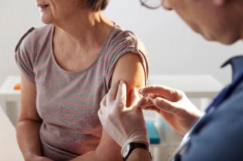
App could improve cancer diagnostics in low- to middle-income and remote areas.
Advertisement
App could improve cancer diagnostics in low- to middle-income and remote areas.
Researchers at Massachusetts General Hospital have developed a smartphone application that can effectively carry out molecular diagnoses in under one hour for an extremely low cost.
The device could enable point-of-care cancer diagnostics for low- to middle-income or remote areas, which often have the highest rates of mortality from cancer due to missed opportunities for treatment.
“In these areas, patient samples often have to be shipped to facilities that are capable of carrying out conventional pathology services,” said Richard Conroy, PhD, program director for Molecular Imaging at NIBIB. “As a result, it can take several days before a diagnosis is returned to the patient. In many cases, patients aren’t able to return for follow-up care either because they have to travel long distances to reach a clinic or can’t afford to take multiple days off work. A low-cost technology that can diagnose cancer at the point-of-care would enable patients to begin treatment on the same day that they are tested, greatly increasing the number of patients who receive treatment.”
The device was developed in part by Hakho Lee, PhD, an associate professor in radiology at Harvard Medical School/Massachusetts General Hospital, and Ralph Weissleder, MD, PhD, director of the Center for Systems Biology at Massachusetts General Hospital.
In the past molecular diagnostics have been difficult to perform at the point-of-care because of the lack of infrastructure and trained personnel, according to Lee.
“To carry out molecular diagnostics currently, you need a good microscope, you need antibodies or ligands that can recognize a molecular target, and you need a specialized person who can interpret the data. Right now, those 3 things are hard to obtain in point-of-care settings,” Lee said.
The new device is called the D3, which stands for digital diffraction diagnosis system. It is made up of a smartphone and an imaging module that attaches to it, comprised of a battery-powered LED light and a lens.
When a sample is collected from a patient in the form of either blood, aspirate, or other biological fluids, it is then mixed with microbeads that have specific antibodies attached to them.
The antibodies then attach to the molecules expressed on the surface of cancer cells and different antibodies are then used depending on the form of cancer detected. The mixture is then placed on a microscope slide and placed into the imaging module, allowing researchers to take pictures of the cell-bead mixture and subsequently provide a diagnosis.
The device is capable of capturing upwards of 100,000 cells per image, which is 100 times the amount of cells that is generally captured with a traditional microscope.
At first, scientists thought they would be able to tell the difference between cancerous and non-cancerous cells by observing which beads stuck to which cells. However, the beads and cell diffracted the light, causing the images to become greatly distorted.
This led researchers to construct an algorithm that reconstructs the images of bead-bound cells from the diffraction patterns that the camera captures.
The reconstruction process requires heavy computations, which made researchers realize early on that they would be limited by the smartphone’s processing capacity.
In order to avoid any delays in the reconstruction process, scientists created an application for the smartphone that automatically uploads the diffraction images as soon as they’ve been taken to a secure cloud that transmits the images to a server at the Massachusetts General Hospital.
The server is able to do many computations at once and reconstructs the images in less than one-tenth of a second.
The server takes the reconstructed images and counts the total number of cells with beads attached to them in addition to the number of beads attached to a given cell. Based on these computations, the cells are classified as high-risk, low-risk, or benign.
The researchers tested the device on a group of 25 patients who had abnormal Pap smear results to test for cervical cancer.
The cell samples were mixed with beads tagged against 3 known cell markers of cervical cancer.
The researchers reported a positive correlation between the number of beads counted and the risk of cancer as confirmed by conventional analysis by a pathologist.
They were able to successfully classify the patients as high-risk or low-risk/benign with 100% sensitivity and 92% specificity.
A pilot study was also conducted by the researchers to determine whether or not they could detect lymphoma cells in fine-needle aspirates of lymph nodes.
The 8-person study showed the device was able to accurately differentiate 4 patients with lymphoma diagnosis from four patients with benign lymph node enlargement.
“The speed at which this technology can diagnose disease is extremely impressive,” Conroy said. “The researchers have taken a process that sometimes takes several days using conventional pathology methods and have condensed it to under an hour. In addition, by taking advantage of cloud-computing and smartphone technology, they’re making the technology available to those who need it the most and for a very low cost.”
In addition to the device’s ability to identify cancer cells, the system also can be adapted to detect DNA. The researchers reported that in their analysis of cervical specimens, they were able to identify the DNA of the human papilloma virus.
This ability to detect DNA opens the door for rapid diagnosis of infectious disease in addition to cancer. Future research by Lee will include expanding the spatial resolution of the images from 2.2 microns down to 1.2 microns through the use of additional computing methods.
In doing this, Lee hopes to allow the device to analyze multiple markers at once by attaching different antibodies to different sized beads and mixing them all together in a sample.
Lee and his team hope to bring the device to Botswana where they will test how easily it can be adapted into local health care systems to detect lymphoma in patients.
Advertisement
Related Content
Advertisement


Latest CME


Advertisement
Advertisement
Trending on Pharmacy Times
1
Pharmacists at the Forefront: Initiating, Monitoring, and Identifying Candidates for VMAT2 Therapy
2
FDA Accepts NDA for AD109 for Treatment of Adults With Obstructive Sleep Apnea
3
Workforce Preparation for Fall Immunization Season Starts Now
4
Could VER-01 Redefine the Role of Cannabis in Chronic Pain Care?
5