
|Articles|December 13, 2010
- December 2010 Heart Health
- Volume 76
- Issue 12
Peripheral Vascular Disease: Increase Awareness to Decrease Risk
Author(s)Jeannette Y. Wick, RPh, MBA, FASCP
Millions of Americans do not know they have peripheral vascular disease, an ofter asymptomatic condition that increases risk of heart attack, stroke, and death.
Advertisement
Millions of Americans do not know they have peripheral vascular disease, an ofter asymptomatic condotion that increases risk of heart attack, stroke, and death.
Increasing awareness of PVD—and its accompanying increases in risk of heart attack, stroke, and death—is imperative.
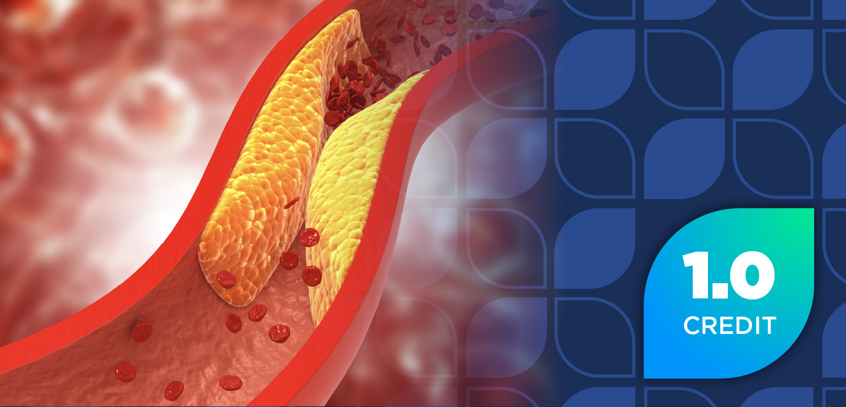
Humans have 3 major vascular beds— coronary, cerebral, and peripheral. Although researchers have studied peripheral vascular disease (PVD; also called arteriosclerosis obliterans) less than diseases in the other 2 vascular beds, PVD’s hallmark symptoms (see Table 1) can cause loss of limb or life.1,2 PVD is an arterial occlusive disease caused by atherosclerotic lesions in the limbs, which diminishes blood supply to the muscles, usually in the lower leg. Although intermittent claudication (IC) is perhaps the most recognized symptom, IC and pain at rest may not develop in sedentary patients who do not challenge their circulation, and PVD progresses steadily. For these patients, the precipitating symptom may be poor wound healing because of inadequate skin perfusion.3
Emanating from atherosclerosis-induced insufficient tissue perfusion, PVD can be acutely exacerbated if emboli or thrombi compromise perfusion. The atherosclerotic process gradually occludes medium and large arteries, more often in the lower extremities than the upper extremities. Emboli tend to lodge at artery bifurcations or in areas where vessels abruptly narrow, and appear to have more severe consequences than thrombi. Thrombi are probably less likely to cause acute and life-threatening symptoms because their slow development allows time for the body to create collateral circulation.4
The greatest risk factors for PVD include smoking, hyperlipidemia, male gender, diabetes mellitus, hyperviscosity, and cancer treatment with radiation or antineoplastic agents. Patients who experience sepsis, hypotension, low cardiac output, aneurysms, aortic dissection, bypass grafts, and underlying atherosclerotic narrowing of the arterial lumen are at increased risk for PVD. The list of risk factors and comorbidities is long, and includes all coronary disease, injury to the vascular bed, and autoimmune disease.5
As noted, PVD can be asymptomatic, as generally symptoms develop slowly and progressively. Intermittent claudication— pain, cramping, or aching in the calf, thigh, or buttocks precipitated by walking and relieved by rest—is often the earliest symptom. With time, collateral circulation may develop, and the patient may find his or her symptoms reduced. Unfortunately, failure to address risk factors and lifestyle often leads to re-emergence. Table 2 describes PVD signs.
Clinicians use the anklebrachial index (ABI) and degree of pallor to determine PVD’s severity. The ABI is obtained by applying blood pressure cuffs to the calf and the upper arm; the systolic ankle pressure is then divided by the systolic brachial pressure. An ABI of 1 or more is normal, and a value less than 0.95 is abnormal.6 ABI is particularly helpful in identifying sedentary patients who are developing PVD, but are still symptomless.
Paralysis or paresthesia may indicate limb-threatening ischemia and should be considered an emergency. As PVD advances, patients’ skin and nails may become shiny or change in texture or pigmentation, or patients may become hairless. In the most advanced stages, patients’ skin may appear mottled in a “fishnet pattern.” The affected area may be numb, cyanotic, cold, and heal poorly. Gangrene is possible. 7
Treatment
Treatment goals for patients who have IC include relieving symptoms and improving exercise performance and daily functional abilities. Usually, physicians prescribe supervised, structured exercise. Patients begin with treadmill or track walking; it usually precipitates claudication, so they rest and try walking again repeatedly for a 30- to 60-minute session at least 3 times a week. Over time, they should improve and experience less pain and cramping.8,9 Physicians also prescribe, as appropriate, pharmacotherapy directed at risk factor modification and antiplatelets to decrease risk of cardiovascular events and improve survival. In some cases, they will consider limb revascularization.
In terms of pharmacotherapy, cilostazol (a phosphodiesterase inhibitor) and pentoxifylline (a hemorheologic methylxanthine) are the only drugs FDA approved for IC. Note that phosphodiesterase inhibitors have caused decreased survival compared with placebo in patients with class III to IV congestive heart failure (CHF), so cilostazol is contraindicated in patients with CHF. More evidence supports the use of cilostazol10- 12 than pentoxifylline, which has been found to be no more effective than placebo in improving treadmill walking distance or functional status in several studies.13-15
Acute limb ischemia—any abrupt decrease in limb perfusion causing a potential threat to limb viability—is diagnosed if symptom duration is less than 2 weeks. Clinicians use laboratory data and imaging to determine if a clot is present. The primary treatment goal is to prevent thrombus propagation and worsening ischemia; usually, heparin is started immediately. Surgery may also be necessary to remove a clot and is used up to 5 times more often than thrombolysis.16 Risk of recurrent limb ischemia is high. Although there are no clear guidelines on duration of anticoagulation, patients will need warfarin prophylaxis for months or years depending on their comorbidities and PVD severity.7
Critical limb ischemia (CLI) is a term applied to patients with typical chronic ischemic rest pain or patients with ischemic skin ulcers or gangrene. It is only used to describe patients who have chronic ischemic disease, defined as the presence of symptoms for more than 2 weeks.
CLI creates a prognosis of high risk for limb loss and for fatal and nonfatal vascular events, myocardial infarction, and stroke. Clinicians should focus on modifying these patients’ cardiovascular risk factors and prescribe antiplatelet drugs. Pedal pain can be intolerable for CLI patients, and limits ambulation severely. Nocturnal ischemic rest pain often wakes patients at night, and leg elevation and cold temperature increase its severity. Consequently, patients may dangle the ischemic leg over the side of the bed or sleep in an armchair.
Pain management is crucial. Ideally, relief of pain is achieved by reperfusing the extremity via surgical revascularization. Prior to surgery, or if surgery is not an option, acetaminophen or nonsteroidal anti-inflammatory drugs can be tried but are rarely effective. Scheduled narcotics are usually required.7,17 Sadly, a large portion of CLI patient care is palliative, as this represents end stage disease.7
Managing patients with CLI ulcers is a multidisciplinary task. When possible, improving circulation can help, as does “offloading”— improving circulation using shoe modifications, orthotics, and casting techniques.18,19 Local infection may occur, and should be treated as an urgent complication with antibiotics and drainage and debridement, if necessary.7
Final Note
Atherosclerosis affects the entire body. Many of the millions of Americans who have PVD are unaware that they have it. Sometimes they are symptomless, and other times they interpret lifestyle changes, like difficulty walking and slow healing, as minor annoyances. Increasing awareness of PVD—and its accompanying increases in risk of heart attack, stroke, and death—is imperative. PT
Ms. Wick is a senior clinical research pharmacist at the National Cancer Institute, National Institutes of Health, Bethesda, Maryland.
Articles in this issue
over 15 years ago
Pharmacy Insights: News & Trendsover 15 years ago
OTC Case Studies: Cardiovascular Disease Conundrumover 15 years ago
Can You Read These Rxs?over 15 years ago
Technology Newsover 15 years ago
Hypertension Watchover 15 years ago
Asthma Watchover 15 years ago
Diabetes Watchover 15 years ago
Women's Health Watchover 15 years ago
Compounding Hotline: Your Compounding Questions AnsweredAdvertisement
Related Content
Advertisement


Latest CME


Advertisement
Advertisement
Trending on Pharmacy Times
1
ADA 2026: Survodutide Cuts Visceral Fat and Normalizes Liver Fat in MASLD. Here's What the Data Mean for Metabolic Risk
2
ADA 2026: Factors Associated With GLP-1 RA Nonresponse in T2D—and What Pharmacists Should Screen For
3
ADA 2026: Optimized Inhaled Insulin Dosing Outperforms Subcutaneous Analogs for Postprandial Glucose in T1D
4
The Hidden Reimbursement Crisis in Medicare’s Drug Price Negotiation Program
5