
|Articles|April 1, 2004
- Volume 0 0
Wound Care in an Aging Population: Special Considerations
Advertisement
Behavioral Objectives
After completing this continuing education article, the pharmacist should be able to:
- Describe the major skin changes related to aging.
- Identify some of the more prevalent wounds in the elderly population.
- Discuss the principles of proper wound care.
- Discuss newer technologies available to consumers that address unique concerns about treating wounds in the elderly population.
According to the 2000 Census, 12.4% of the US population, or 35 million people, were 65 years of age or older. The trend was smaller than reported in 1990, but it is estimated to be temporary, as the babyboomer population reaches age 65 beginning in 2011. People 85 years and older are the fastest-growing age group within the older population. The opportunities to care for and interact with the elderly are vast and unavoidable in all pharmacy practice settings.
Many intrinsic and extrinsic variables contribute to the aging of society, and improved health care has increased life expectancy. There still exist, however, increases in incidences of morbidity, nonthreatening diseases, and chronic illnesses that require health and social care services.
This article will focus on special considerations needed when providing wound care to the elderly. Approximately 3.5% of the geriatric population suffers from leg ulcers and, because this number rises as the population ages, recurrence rates are as high as 70%. The increased prevalence of wounds in the elderly may be caused by immobility, disease, or merely changes in the skin itself.
Today pharmacists are in a unique position to deliver wound care in a number of settings as part of the multidisciplinary team of health professionals. To do so, members of the profession must ensure a high standard of practice and a constant updating of skill. Understanding the natural aging process of the skin and the healing process allows pharmacists to better advise and care for geriatric patients who present with wound care issues.
Structure of the Skin
The epidermis, the outermost layer of the skin, is composed of basal cells, keratinocytes, Langerhans' cells, melanocytes, and stratum corneum. This avascular layer provides protection against environmental toxins and acts as a barrier to prevent essential bodyfluid loss. The basal cells aid in epidermal production. Keratinocytes provide a waterproof covering, and Langerhans' cells provide antigen presentation. Melanocytes synthesize melanin, which determines the skin's color and protects the skin from sunburn and ultraviolet radiation.
The dermis, the middle layer of the skin, is composed of blood vessels, collagen, reticulum, elastin, fibroblasts, lymphatic glands, macrophages, mast cells, and nerve fibers. Within this layer, thermoregulation is maintained by vascular structures, and skin proteins provide skin elasticity, strength, and texture. Healthy dermal components are vital for wound repair and healing, in terms of blood circulation, inflammatory response, foreign-substance phagocytosis, and proper lymphatic drainage.
The innermost layer of the skin, the subcutaneous tissue, is composed of adipose, connective tissue, blood vessels, nerves, and lymphatic vessels, and it serves 2 main functions. Adipose provides thermoregulation of energy storage and balance by limiting conductive heat loss. The subcutaneous layer also protects the body from shear-related injury, shock, and trauma.
Structural Skin Changes Associated with "Normal" Aging
The natural aging process is an inevitable decline of the body's organs, including the skin. When a person is in his or her 20s, subtle visible effects and physiologic skin alterations begin, regardless of external environmental exposure. Between 30 and 80 years of age, the epidermis becomes thin, as the turnover rate of the structural cells decreases by approximately 30% to 50%. This decrease compromises nutrient transfer and communication between the 2 layers, affecting barrier, immunologic, and mechanical functions. The decrease in melanocytes (loss of 10%- 20% per decade) and in Langerhans' cells increases the risk of skin cancer and infection.
The interface between the epidermis and the dermis flattens, and the loss of integrity increases the skin's vulnerability to simple trauma and to shearing forces that may cause tearing and blistering. There is a loss of dermal thickness in 20% of the elderly as the dermis becomes avascular. Degradation and dysregulation of collagen and collagen synthesis impair the skin's strength and ability to heal properly. There is a progressive loss of elastic recovery of the skin.
The sites of fat alterations can limit the main functions of the subcutaneous layer of skin. Although the proportion of subcutaneous fat actually increases until age 70, the overall volume of fat diminishes with age. Thinning of subcutaneous tissue in the face, hands, shins, and feet causes the skin to become dry, flaky, and itchy and decreases insulation, padding, and protection against mechanical injury. Pressure sores over bony prominences often occur in the elderly, due to the skin's inability to diffuse pressure over these areas.
Over time, aging skin begins to sag and wrinkle. It is increasingly inelastic, rigid, and unresponsive to stress and wound healing. Proper treatment and care must be taken to prevent excessive damage or injury to an aging person's integumentary system. If it is damaged or compromised, skin lesions and wounds can create medical challenges in treatment and quality-of-life issues for the elderly.
General Principles of Wound Healing
Any break in the skin is considered a wound or sore, regardless of the acuity of the break. There are 3 distinct biological phases in the wound-healing process of restoring cellular structure and tissue layers: inflammation, proliferation, and remodeling or maturation. All 3 phases are integral to the completion of the healing process, and they may occur at various rates. Scar formation may occur from excessive fibroblastic proliferation or inadequate healing.
Inflammation Phase
In this early defensive or reaction phase, vascular and cellular response to an injury occurs. Epinephrine, norepinephrine, prostaglandins, serotonin, and thromboxane mediate a short vasoconstriction response. This response causes a temporary blanching of the wound area to reduce hemorrhage, increase platelet aggregation, and retain healing factors within the wound. The intrinsic and extrinsic coagulation cascades initiate activation of thrombin, which converts fibrinogen to fibrin. Fibrin is the essential primary component of the wound matrix to which inflammatory cells, platelets, and plasma protein migrate. Wound healing is delayed or impeded if the fibrin matrix is removed. Vascular permeability is facilitated by the migration of inflammatory cells and factors to the site of the injury.
After the brief period of vasoconstriction, there is a vasodilation period mediated by histamine, kinins, leukotrienes, and prostaglandins. The wound site becomes edematous, erythematous, and heated because of increased blood flow and debridement of the wound by infectionfighting cells. Pain may be experienced due to tissue and bacterial degradation, alterations in pH, tissue swelling, and hypoxemia.
The most significant cells involved in the healing process during the inflammatory phase are macrophages. They rid the injury site of debris, as well as secreting growth factors essential in the formation of new tissue and blood vessels (angiogenesis). If macrophage involvement is impaired, the overall healing rate is delayed.
Proliferation Phase
The proliferative phase?also called the connective tissue, fibroblastic, or regeneration phase?consists of 4 stages: epithelialization, fibroplasia, angiogenesis, and contraction. This phase may last 4 to 24 days after the injury. The main event during this phase is the formation (3-5 days after the injury) of granulation tissue that is comprised of collagen, a fibronectin matrix, glycoaminoglycans, and proteoglycans. Granulation-tissue formation is initiated during the earlier inflammatory phase and overlaps into the proliferative phase.
Epithelialization is the formation of epithelium over an exposed surface. This process can occur within 24 to 48 hours of the injury, and the new layer provides a barrier between the external environment and underlying viable tissue. Adequate tissue humidity will expedite epithelialization and can be ensured with the application of occlusive and semiocclusive dressings within the first 48 hours after the injury. A scar forms when epithelialization is complete.
Fibroplasia begins 3 to 5 days after the injury and may continue for up to 14 days. Fibroblasts are vital to granulation tissue and are responsible for the synthesis of collagen. Collagen aids in stabilizing the fibronectin matrix and is the main component of the final scar. Elastin, present in small quantities, contributes to the skin's recoil and stretch properties.
Proper blood flow is critical to proper wound healing. Macrophages stimulate angiogenesis that increases perfusion of healing factors to the wound. Once the demand for new blood vessels decreases, angiogenesis ceases.
Centripetal movement of wound edges or contraction occurs simultaneously with collagen production 5 to 15 days after the injury. The rate of contraction is dependent on the shape of the wound and the tolerance of tissue. Square wounds and loose tissues contract more than round wounds and tissues with poor tolerance.
Maturation Phase
Collagen and cytokines are required during the maturation phase, as collagen remodeling begins approximately 21 days after the injury and may continue indefinitely. During collagen remodeling, the tensile strength of the wound recovers at a gradual rate, reaching 20% at 30 days, 70% at 6 months, yet never fully recovering to the strength of unwounded skin. Cytokines are mediators in the healing process that bind to cell-surface receptors to stimulate cell responses, and they are involved throughout all phases of the healing process.
Factors That May Impede Wound Healing
Pharmacists can educate patients about local and systemic factors that impede wound healing. Local factors include dry environment, edema, incontinence, infection, necrosis, and pressure. Systemic factors include age, body build, chronic diseases, nutritional status, vascular insufficiencies, and radiation and immunosuppression therapy.
Local Factors
Exposure to the air reduces the surface temperature of the wound and further delays healing. Wounds heal 3 to 5 times faster in a moist environment than in a dry environment. Moisture-retentive dressings keep the wound moist and enhance epithelialization. Edema interferes with cellular nutrition and with bringing oxygen to the wound, and diuretics often are used to decrease fluid retention. Fecal and urine incontinence can alter the skin's integrity, increasing the risk of injury as well as impaired wound healing. Local or systemic infection delays healing, and the infected area should be cultured or assessed for appropriate antibiotic therapy, or to rule out osteomyelitis if there is bone involvement. Dead tissue does not regenerate and should be removed before healing can occur. Necrotic tissue may present as slough (wet, stringy, loose, and yellow tissue) or eschar (dry, leathery, and dark or black tissue). Peripheral vasoconstriction or excessive or continuous pressure on the wound site can impair proper blood flow, as well as impairing the supply of oxygen, nutrition, and other factors to the surrounding tissue. Tight stockings or leg coverings or bandages wrapped too tightly over a wound may cause these problems.
Systemic Factors
Geriatric patients generally heal more slowly than younger patients. Skin breakdown and delayed wound healing may result from inadequate nutrition intake, dehydration, and compromised states. Body build can impair wound healing, because excessive adipose tissue impairs blood flow and a thin build (due to the lack of nutrition and oxygen) also delays healing. Wounds in patients with diabetes often heal more slowly because of the need for insulin in granulation formation. Arterial and venous insufficiencies impair blood supply to tissues surrounding wounds. Radiation therapy can alter skin structure and cause ulcerations. Immunosuppression from disease or drug therapy delays wound healing.
Patients have less control over systemic factors than over local factors that impede wound healing. Pharmacists can educate and counsel patients to identify problems, can encourage compliance with drug therapy, and can promote lifestyle behaviors to decrease the risk of impairment and to increase successful wound healing.
Types of Wounds
Wounds in an aging population are common problems, particularly leg ulcers, pressure sores, and skin tears. Types of wounds common in the elderly include the following:
Chronic Wounds
- Leg ulcers
- Mechanical stress wounds
- Diabetic wounds
Acute Wounds
- Lacerations
- Grazes
- Burns
- Skin tears
Leg Ulcers
Leg ulcers have a number of different causes, including venous insufficiency, arterial disease, diabetes mellitus, vascular complication of autoimmune disease (such as rheumatoid arthritis), malignant disease, trauma, and deliberate self-injury (Table 1). Leg ulcers, more than any other chronic wounds, present a diagnostic challenge when they are seen in the pharmacy.
Venous Ulcers
These ulcers result from venous hypertension, due mainly to a breakdown in venous circulation in the leg via the calf muscle pump. This breakdown is associated with an inability to force the passage of venous blood from the superficial veins and through the perforating veins to the deep vein system. Venous insufficiency develops as a result of obstruction or valvular incompetency. Throughout the venous system, bicuspid valves control the flow of blood pumped with the aid of muscular contraction, and malfunction may be due to deep vein thrombosis (DVT), abnormalities, and degeneration. Valvular malfunction causes the reflux of blood back through the superficial systems, producing hypertension and varicosity. Venous ulcers have certain general features (Table 2).
Deep vein thrombosis is a common cause of venous ulcers. It is often undiagnosed at the time of hospitalization for surgery or fracture (silent DVT). Obesity and decreased mobility in people who stand on their feet for long periods of time can cause venous stasis. A family history of varicose veins also increases the risk for developing venous ulcers.
The single most important aspect of the management of simple venous ulceration is the application of adequate levels of graduated compression, either as stockings or bandages. It is important to note, however, that antiembolic stockings are not appropriate in the management of varicose veins, because they do not deliver sufficient compression.
Arterial Ulcers
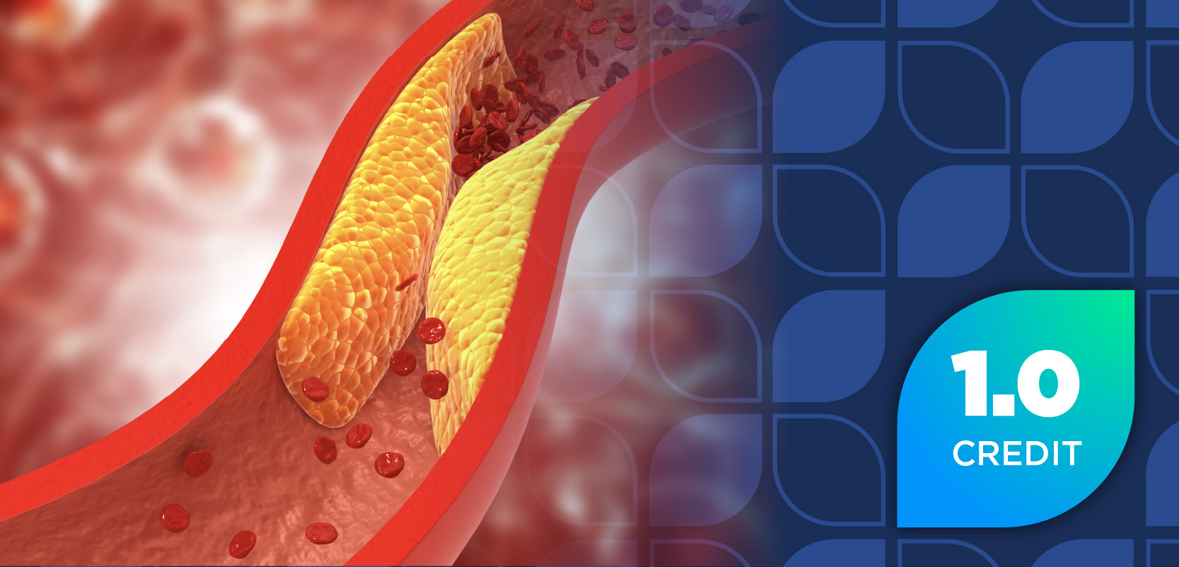
Arterial ulcers result from a lack of circulation through the microcirculatory or main arterial system. The lack of blood flow starves the wound of necessary oxygen, nutrients, and important healing factors that are present in arterial blood. The main cause of arterial vessel malfunction is extramural or external strangulation of an artery that obstructs blood flow due to scar tissue, fibrosis, edema, chronic infection, or neoplasm. Mural or vessel wall changes, atherosclerosis, or plaque formation also slows the rate of blood flow through the arteries. Intramural changes or the blockage of vessels can be caused by thrombosis or embolism. Arterial ulcers have certain general features (Table 3).
Arterial ulcers often are seen in patients with uncontrolled diabetes. Smoking is a significant factor in the reduction of arterial blood flow, due to increased platelet adhesiveness caused by nicotine, reduced oxygen at the wound caused by carbon monoxide, and reduced protein replication and enzyme inhibition caused by hydrogen cyanide. A history of peripheral vascular disease is a common cofactor.
Other Causes of Leg Ulcers
Trauma is an initial cause of skin break that may develop into an ulcer, often due to other complications, such as underlying poor health and skin condition. This trauma also may result from poorly applied compression bandages.
Vasculitic ulcers may develop in association with autoimmune diseases, such as rheumatoid arthritis and polyarteritis. Infections of the skin can produce ulcers, especially from the necrotizing types of bacteria associated with insect bites. Some ulcers result from neoplasia, and neoplasia may develop in some nonhealing ulcers. The most common carcinomas are squamous cell and basal cell.
Patients with diabetes develop leg and foot ulcers, mainly arterial in nature, as a result of reduced blood flow in the extremities. They also may develop neuropathies: ones that cause a loss of feeling in the feet; motor neuropathies, in which there is a change in bone and tendon structure; or autonomic neuropathies, which cause a change in skin tone. The diabetic patient is at greater risk of ulcer formation because of these neuropathies and has a fivefold greater risk of infection, compared with the nondiabetic patient. Patients with diabetes also have a reduced inflammatory response and may suffer from irreversible nerve damage.
Ulcers also may develop from lymphedema caused by reduced function of the lymph vessels to drain extracellular fluid. Although many ulcers look similar on first view, a closer examination of both the wound itself and the patient's history will help provide a guide to the possible cause of the wound. It may be necessary to refer the patient for diagnostic tests to clearly establish the underlying cause. Once the cause is identified, the most appropriate treatment can be commenced.
Mechanical Stress Wounds
Pressure ulcers are the most preventable of all chronic wounds. They may be simple blisters from ill-fitting footwear, or extensive pressure sores experienced by patients bedridden due to stroke, spinal injury, multiple sclerosis, or dementia.
It has been estimated that 6% to 12% of all patients treated in hospitals develop a pressure wound, but, sadly, this number increases to ~30% in the elderly. A pressure wound develops when the capillary blood flow to the skin and tissue over a bony prominence is decreased for a sufficient period of time.
The capillary pressure in the arterial blood system is usually 32 mm Hg. A pressure of only 30 mm Hg restricts arterial blood flow. The consequence of this restricted blood supply is a reduction in oxygen supply and nutrition to the tissue, accompanied by the problem of waste products not being removed from the site.
The results of reduced blood flow are hypoxia, tissue acidosis, and increased capillary permeability, which allows intravascular fluid to escape, causing edema and cell death. Pressure wounds are commonly caused by direct pressure, friction, and shear forces.
Pressure Wounds
Direct pressure on tissue over a bony prominence in excess of 30 mm Hg will cause ischemia in the surrounding tissue. This pressure can occur when a patient is lying in a bed or gurney or sitting in a chair. The extent of tissue damage depends on the intensity of the pressure and the length of time the pressure remains unrelieved. Tissue tolerates pressure for short periods of time; however, even low pressure over a long period of time will have some detrimental effect.
Friction Wounds
Friction wounds occur when the top layers of skin are worn away by continued rubbing against an external surface. This type of wound may be a simple blister or tissue edema or an open pressure wound. It is often caused by ill-fitting footwear or wrinkled bed linen.
Shearing-Force Wounds
Shearing-force wounds occur when skin is unable to move against a surface while underlying bone and tissue are forced to move beneath the skin. Shearing force contributes to the destruction of microvasculature in a manner similar to direct pressure. This type of pressure injury is seen in patients who remain sitting up in bed or in a chair, while gravity causes them to slide down and their skin adheres to the bed linen or the surface of the chair.
Acute Wounds
The major acute wounds in the aging population are skin tears. The main causative factor is trauma from the following:
- Manual handling (eg, transferring the patient from bed to chair)
- Removal of adhesive tapes
- Falls
- Bed rails, cot sides, or wheelchair foot plates
General Principles of Wound Care
All wounds heal by the same process; however, the treatment of a wound depends on its severity and size. The goals of wound care are to keep the wound moist, clean, and free from infection and trauma, as well as to maintain the skin's integrity. Acute wounds usually are abrasions, burns, lacerations, or punctures that are limited and localized to the site of the injury. If optimal care is provided, these wounds will heal within 4 to 14 days with minimal scarring.
Minor abrasions and scrapes usually require only washing the area with mild soap and warm water as soon as possible after the injury and keeping the area protected with a sterile bandage. For these types of acute wounds, an initial thorough cleansing is recommended. Then the wound should be kept covered, with a fresh bandage as needed. Washing acute wounds more than once is not recommended unless there is suspicion of contamination. Each additional washing will disturb the healing wound bed.
Ideally, a liquid detergent or washing solution with surfactant action (eg, QV Wash) should be used, which has a lower pH of 5 or 6 than soap (pH 9-12) and is less irritating to an open wound. Bleeding should be minimized by covering the wound area with a clean dressing and applying pressure for 10 minutes. Dirt and debris should be removed from the wound with water pressure under a briskly running faucet or shower nozzle. If the wound is dirty, and if it has been 5 years since the patient's last tetanus immunization, another shot should be received.
Medical attention is needed if any of the following is true:
- A tetanus shot is needed
- The wound is longer than 1/2 inch
- The wound is deep to the fat tissue or bone
- Bleeding does not cease after 10 minutes of pressure
- Dirt or debris cannot be removed without scrubbing
- The wound is not healing
- Redness extends from the wound after 2 days
- Pus or a yellow discharge is present
Chronic wounds, ulcerations, or acute wounds that do not heal well often require a multidisciplinary approach with several treatment modalities. Procedures may include culturing the wound to identify contaminating organisms; irrigation; debridement (autolytic, enzymatic, or mechanical); wound dressings; and support surface therapy.
Wound Treatments
From the earliest practices of pharmacy, treating injuries and wounds has been a daily role for members of the profession. Over the past 80 years in particular, pharmacists have played a significant part in the development of wound products. The choice of product will depend on the wound type, the wound color, the wound depth, and the level and type of exudate.
The traditional theories have been that a wound should be kept clean and dry so that a scab may form over it; that a wound should be exposed to air and sunlight as much as possible; and that where tissue loss is present the wound should be packed to prevent surface closure before the cavity is filled, and then the wound should be covered with a dry dressing.
Clear disadvantages of these principles are that a scab composed of dehydrated exudate is a physical barrier that delays healing, because epidermal cells cannot move easily under the scab, and there may be poor cosmetic results and scarring. When a wound is packed with dry gauze, the quality of healing is impaired due to adhesion of the material to the wound surface, which causes the wound to dry out.
Indeed, for many years, the products used were "passive" or employed the "plug and conceal" concept. These products included absorbent cotton wool gauzes and lint. Such products, especially gauze, have no real place in modern wound management. Gauze, as a primary dressing, in addition to causing the wound to dry out, is easily penetrated by bacteria. It also increases the risk for contaminating the wound when gauze fibers break off and fall into the wound. Gauze adheres to the wound and tends to traumatize the wound when it is removed.
Wounds covered by an occlusive dressing do not form a scab. Epidermal cells are allowed to move rapidly over the dermis surface through exudate, which collects at the wound/dressing interface. The application of a totally occlusive or semipermeable dressing to wounds also prevents secondary damage as a result of dehydration.
Classification of Wound Dressing Products
Inert Products
Inert products include gauze, lint, and cotton dressings. Other simple dressings include nonadherent dressings, which are useful over minor lacerations or as a protective dressing once a wound has healed.
Tulle gras, or paraffin gauze dressing, retains moisture by producing a waterproof paraffin cover over the wound. The use of this dressing may lead to maceration caused by water vapor and exudate that cannot pass through the paraffin, which are therefore trapped within the wound itself. Tulle gras also is permeable to the air and bacteria and has been known to adhere to a wound, risking trauma on removal. It has minimal use for treating simple grazes and first-degree burns and has been used as an initial dressing over skin grafts.
Newer alternative tulle-type dressings are composed of tightly woven, synthetic fibers (not gauze). They have a base material that allows moisture to pass through and minimizes maceration to the wound and surrounding tissues.
In addition to gauze and lint, there are other simple, modified absorbent pads covered with a perforated plastic film to prevent adherence to a wound, which are used as both primary and secondary dressings. These pads are used in minor wounds and wounds with low exudation.
Interactive Dressings
Interactive dressings use the environment provided by the body itself to encourage normal healing.
Semipermeable Films. Semipermeable dressings consist of a thin, polyurethane membrane coated with a layer of acrylic adhesive. Their physical properties include being gas- and water vapor?permeable, impermeable to microorganisms, and transparent and flexible. Wound assessment is easy, and the dressings are conformable, offering considerable patient comfort. They provide a moist environment and encourage autolysis. Semipermeable films, however, do not have the ability to absorb exudate. They are particularly useful on superficial, clean wounds and in the prevention of breakdown and pre-ulcers in pressure wounds. They should not be used on infected wounds. There are no absolute contraindications for their use; however, some local irritation has been observed from the adhesive.
Polyurethane Foams. These products meet many standard requirements for an ideal dressing, in that they allow exudate to pass through the nonadherent surface, to be absorbed in the main compartment of the product, while maintaining a moist and insulating environment. They may be composed of soft, open-cell, hydrophobic foam, with a heat-treated contact layer that produces a nonadherent hydrophilic layer. Some foams are soft and hydrophilic, bonded to a semipermeable polyurethane film and covered with a 3-dimensional plastic net.
These products are highly absorbent of exudate and maintain a moist environment without maceration of surrounding tissue. Polyurethane foams are used mainly in moderate to heavily exudating wounds?including ulcers, donor sites, and minor burns?and they act as a secondary dressing, particularly as a covering for use with amorphous hydrogels. There are no known contraindications; however, they have little value in low-exudating or dry wounds with eschar. They come in a variety of sizes, including shapedcavity devices, which may be inserted into pressure wounds or dehisced surgical wounds.
Hydrogels. Hydrogels are a group of complex organic polymers that have high water content (30%-90%). This broad class of polymers swells extensively in water but does not dissolve in water. They have properties of both rehydrating dry tissue and absorbing certain amounts of fluid. Hydrogels as amorphous gels help rehydrate "sloughy" wounds and necrotic tissue. They also aid in autolytic debridement of wounds and in the management of burns, including sunburn, scalds, and other partial-thickness burns. In addition, amorphous hydrogels have been used in the management of chickenpox and shingles, applied liberally to the eruptions 4 to 5 times daily to provide a moist environment, relieve discomfort from lesions, and reduce the probability of scarring.
Hydrogels also are available in a sheet form that consists of a crosslinked polymer hydrogel and water held in a backing. These products are particularly useful in the management of burns and aid in the removal of necrotic tissue in pressure wounds.
Bioactive Dressings
Bioactive dressings utilize the environment provided by the body. They also stimulate the healing cascade.
Alginates. Alginates are calcium or sodium/calcium salts of alginate acid, composed of mannuronic and guluronic acids obtained from seaweed?mainly from the genus Laminaria. They are practically insoluble in water and organic solvents. When applied to a wound, sodium salts present in the wound exchange with calcium in the alginate to form sodium alginate, a hydrophilic gel. This gel has the ability to absorb exudate while maintaining a moist environment at the interface between the dressing and the wound. Alginates rich in guluronic acids form strong but brittle gels, whereas alginates rich in mannuronic acid are weaker but more flexible gels.
The physical properties of alginates include the ability to absorb exudate to form a gel, and to produce a moist environment without maceration of surrounding tissue. Some have hemostatic properties.
Alginates are used on donor sites, bleeding sites, exudating leg ulcers, and cavities. There are no known contraindications; however, they have little value in low-exudating or dry wounds with eschar. They come in a number of different forms, including sheets and packing rope, and in combination with charcoal for exudating malodorous wounds.
Fiber dressings, or hydrofibers, are composed of carboxymethylcellulose sodium, which absorbs exudates and forms a firm gel. These dressings differ from alginates in that they do not wick laterally and may be placed in the wound and/or around it to protect the peri-skin from maceration.
Hydrocolloids. Hydrocolloids are a combination of polymers held in a fine suspension and often contain polysaccharides, proteins, and an adhesive. When placed on a wound, polymers combine with exudate and form a soft, moist gel-like mass. They also encourage autolysis to aid in the removal of slough from a wound. Hydrocolloids were originally introduced as stoma adhesive to protect good skin around stomas. It was subsequently found that they improve the healing of excoriated skin.
The early forms of hydrocolloids are occlusive and thus impermeable to gases and water vapor. They do, however, act as a barrier to external bacteria and are waterproof, so that the patient can bathe. Thin, transparent hydrocolloids have a polyurethane film backing, are nonocclusive, and do allow passage of water vapor and gases.
Hydrocolloid products are used in low to moderately exudating wounds?including ulcers, donor sites (after hemostasis), and granulating wounds?and they act as secondary dressings. They may be used in conjunction with hydrocolloid paste or powder in deep ulcers or cavities. They convert to the same hydrophilic gel and are covered with the normal hydrocolloid wafer.
There are known sensitivities to these products, mainly to the adhesives. They are contraindicated in dirty and infected wounds and in deep wounds with involvement of muscle, tendon, or bone.
Hydrocolloids are available in a thin form, which is permeable, very lightweight, and comfortable. They are used over sutured wounds and incision sites as protective dressings, as well as on lightly exudating wounds, because they have the ability to cope with exudates.
Hydroactive Dressings. Hydroactive dressings are composed of absorbent polymers that absorb exudate and expand while maintaining a moist environment and aiding in autolysis without maceration of the surrounding tissue. They are used on moderate to heavily exudating wounds, including ulcers and granulating wounds.
Some hydroactive dressings are confused with foam dressings. They appear to be similar, but foams absorb by siphon and hold exudates in air spaces, and hydroactive dressings absorb exudates into a polymer structure and expand in shape. These dressings are available in thin, nonadhesive cavity and island forms.
Miscellaneous Dressings
There are a few important new dressings that do not fit into the previous categories of dressings. In recent years, new dressings have been developed that incorporate active silver into the structure. They are used in both infected and critically colonized wounds to lower bacterial burden in the wound and to prepare the wound bed for healing.
Silver dressings come in a number of forms, including foam with silver, hydrocolloid with silver, hydroactive with silver, alginate with silver, hydrofiber with silver, and hanacrystalline silver in a simple base. The choice of silver dressing depends on the wound type, the depth and level of exudation, and the bacterial burden.
The bacterial burden also may be reduced by the use of newer forms of iodine. Cadexomer iodine is a complex composed of a polysaccharide polymer with iodine cross-linked into its structure. Exudates in the wound cause iodine to be released over a 72-hour period, reducing bacterial levels in the wound. Simultaneous absorption of exudates has been shown to stimulate certain growth factors in the wound. This product is available as a paste, a powder, and a backed dressing and is used in diabetic, recalcitrant nonhealing, sloughy, and malodorous wounds.
Team Approach to Wound Care
If wound healing is not successful or progresses to a chronic state, patients can be referred to wound care centers, which are staffed with a variety of health care professionals, including physicians, nurses, pharmacists, physical therapists, and dietitians. Chronic-wound healing can be challenging to patients and caregivers. A team approach with patients and professionals can facilitate a positive relationship that will increase the healing rate of chronic wounds. Pharmacists are accessible resources to identify patients who need medical attention and to provide advice to patients in wound care products selection.
Wound Care in an Aging Population: Special Considerations
This educational lesson will be available to pharmacists on-line at www.pharmacytimes.com.
(Based on the article starting on page 76.) Choose the 1 most correct answer.
1. Which of the following statements about the structure of skin is true?
- Adipose in the epidermis layer provides thermoregulation.
- Melanocytes in the dermis aid in protection from sunburn.
- Skin proteins in the dermis provide tensile strength and elasticity.
- Vascular structures in the epidermis provide healing factors to wound sites.
2. When a person ages, the following structural change in the skin can be seen:
- Loss of dermal thickness results as the dermis becomes avascular.
- The overall volume of fat distribution in the body increases with age.
- Infection risk increases as the number of Langerhans' cells increases.
- The turnover rate of cells decreases, resulting in subcutaneous thinning.
3. Which of the following is not a phase in the wound-healing process?
- Maturation
- Dehiscense
- Proliferation
- Inflammation
4. The main function of fibroblasts is to:
- Initiate scarring.
- Synthesize collagen.
- Initiate inflammation and repair.
- Provide metabolic skin requirements.
5. During the proliferative phase:
- Angiogenesis is stimulated by macrophages.
- Contraction occurs from decreased collagen production.
- Cytokine-mediated cell responses form a fibronectin matrix.
- Vasodilation occurs as healing factors migrate toward the wound.
6. SK is a 79-year-old man with type 2 diabetes mellitus, lung cancer s/p chemotherapy, and a Stage III decubitus ulcer. He comes to the wound care center for consultation. Which of the following is an extrinsic factor that may affect his healing process?
- Age
- Chemotherapy
- Diabetes mellitus
- Inappropriate wound care
7. Which of the following statements is true regarding factors that impede healing?
- Eschar tissue is seen as wet, stringy, and loose tissue.
- Incontinence interferes with cellular nutrition to the wound.
- Dry environments are important to decrease bacterial growth.
- Excessive adipose tissue impairs oxygen delivery to the wound.
8. Which of the following is a characteristic of a venous ulcer?
- Painful
- Defined border
- Irregular shape
- Shiny appearance
9. Which of the following is a characteristic of an arterial ulcer?
- Dusky foot
- Edematous
- Varicose veins
- Eczema present
10. All of the following are associated causes of venous ulcers except:
- Obesity.
- Smoking.
- Deep vein thrombosis.
- Valvular incompetence.
11. All of the following statements are goals of wound care except:
- Maintain skin's integrity.
- Prevent scarring.
- Prevent infection.
- Keep wound dry.
12. Which of the following is an example of an inert wound care product?
- Paraffin gauze
- Silver dressing
- Polyurethane foam
- Semipermeable film
13. Which of the following statements is true regarding tulle gras?
- It is waterproof.
- It does not adhere to the wound.
- It is impermeable to bacteria.
- It is preferred for use in firstdegree burns.
14. All of the following statements are true regarding semipermeable films except:
- They maintain a moist environment.
- They facilitate autolysis.
- Local irritation may occur from the adhesive.
- They are recommended for use in infected wounds.
15. Which of the following dressings is available in a shaped-cavity device for insertion into wounds?
- Alginates
- Hydrogels
- Polyurethane foams
- Silver dressing
16. This type of dressing is particularly useful in the management of severe burns.
- Alginates
- Hydrogels
- Polyurethane foams
- Semipermeable films
17. This type of dressing is contraindicated in infected wounds.
- Alginates
- Hydrocolloids
- Paraffin gauze
- Polyurethane foams
18. Autolysis is encouraged by which of the following types of dressing?
- Alginates
- Cadexomer iodine
- Hydrocolloids
- Tulle gras
19. This type of dressing will not absorb exudate from wounds.
- Hydrogels
- Hydrocolloids
- Polyurethane foams
- Semipermeable films
20. This dressing has been used to relieve discomfort from chickenpox and shingles lesions.
- Alginates
- Cadexomer iodine
- Hydrogels
- Silver dressing
CE ANSWER FORM INSTRUCTIONS
TESTING AND GRADING PROCEDURES NEW SCORING OPTIONS
- Each participant achieving a passing grade of 70% or higher on any examination will receive a statement of credit stating the number of CE credits earned. This form should be safeguarded and may be used as documentation of credits earned.
- Participants receiving a failing grade on any exam will be notified and permitted to take 1 reexamination at no extra cost.
- All answers should be recorded on the answer form attached. For each question, decide which choice is the best answer, and circle the letter of the response representing your choice.
- Mail your completed exam form to the following address: Pharmacy Times, 405 Glenn Drive, Suite 4, Sterling, VA 20164-4432.
NEW SCORING OPTIONS
- Fax: 703-404-1801
- This lesson is FREE on-line, receive instant grading, as well as download your certificate? www.pharmacytimes.com
Please print clearly?certificate will be issued from information given.
Please mail completed forms with your payment to:
Pharmacy Times CE Department, 405 Glenn Drive, Suite 4, Sterling, VA 20164-4432
Articles in this issue
about 22 years ago
RxPRODUCT NEWS PROFILE: Caduetabout 22 years ago
COMPOUNDING HOTLINEabout 22 years ago
Case Studiesabout 22 years ago
Buddies Are Good for the Heartabout 22 years ago
Low-Fat Diet Shows Promise for Prostate Cancerabout 22 years ago
Rage Is Connected to Stroke Riskabout 22 years ago
InnoLetabout 22 years ago
Colace/Peri-Colaceabout 22 years ago
TampAlerTabout 22 years ago
Low Testosterone Is Linked with Alzheimer's DiseaseAdvertisement
Related Content
Advertisement

Latest CME


Advertisement
Advertisement
Trending on Pharmacy Times
1
ADA 2026: Factors Associated With GLP-1 RA Nonresponse in T2D—and What Pharmacists Should Screen For
2
ADA 2026: Survodutide Cuts Visceral Fat and Normalizes Liver Fat in MASLD. Here's What the Data Mean for Metabolic Risk
3
ADA 2026: Optimized Inhaled Insulin Dosing Outperforms Subcutaneous Analogs for Postprandial Glucose in T1D
4
The Hidden Reimbursement Crisis in Medicare’s Drug Price Negotiation Program
5