
|Articles|July 11, 2011
- July 2011 Digestive Health
- Volume 77
- Issue 7
Understanding Gallstone Disease
Author(s)Guido R. Zanni, PhD
Gallstone disease is often asymptomatic, and health care professionals are divided over whether these patients should wait to seek treatment.
Advertisement
Gallstone disease is often asymptomatic, and health care professionals are divided over whether these patients should wait to seek treatment.
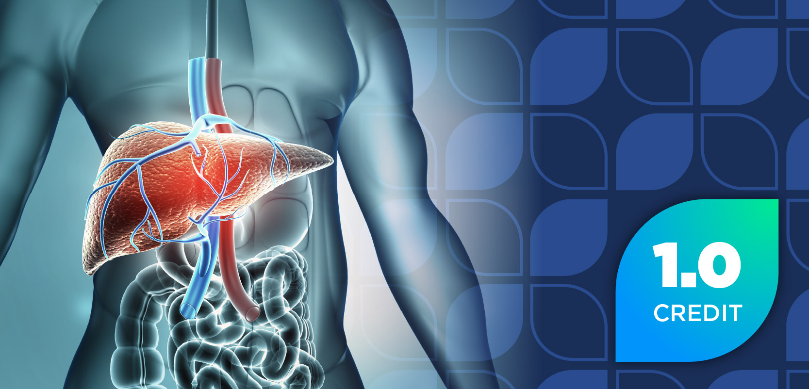
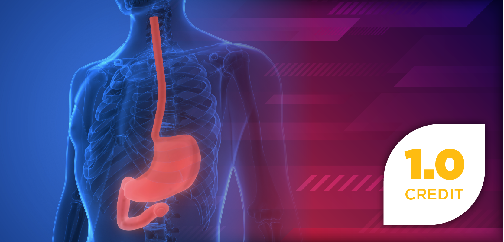
Shaped like a small pear, the gallbladder sits in the upper right side of the abdomen. Its primary function is to store bile produced by the liver, which will be later released into the small intestine to facilitate digestion, especially the absorption of fats.1 Gallstone disease—the crystallization of bile salts or cholesterol in the gallbladder—affects 10% to 15% of Americans, with approximately 1 million cases diagnosed each year.2
Gallstones occur in all age groups, but the incidence increases with age. Risk factors include obesity, rapid weight loss (more than 3 lb a week), parenteral nutrition, pregnancy, the use of contraceptives or estrogen replacement therapy, diabetes, use of cholesterol-lowering agents, and heredity (25% phenotype variance seen in twins).3,4 Ethnicity is also a risk factor, with incidence nearly 100% in Native Indians of Peru and Chile.5 Closer to home, increased incidence is found in American Indians and Mexican Americans.4
Cholelithiasis (gallstone formation) results from a combination of factors, including supersaturation of cholesterol and bile, resulting in crystallization of cholesterol;
acceleration of nucleation of cholesterol monohydrate in bile (molecules begin to crystallize and form nuclei, which then exponentially attract other molecules to crystallize); bile stasis; and delayed gallbladder emptying.6
Gallstones are categorized by their composition.
• Cholesterol stones, yellowish in color, are composed of undissolved cholesterol and account for up to 80% of observed stones
• Pigment stones, dark brown or black in color, result from increased bilirubin production
• Mixed stones contain both bilirubin and anywhere from 50% to 90% cholesterol4-6
Gallstones range from microscopic, also called biliary sludge, to more than an inch in size.2 Studies demonstrate that small stones are more serious, because they can lead to pancreatitis.3
Gallstone Complications
Approximately 90% of patients with gallstones are asymptomatic; their stones are often detected during diagnostics for another condition. For the remaining 10%, the most common complications are biliary colic, which is sharp, intense pain lasting up to several hours due to the forceful contraction of the gallbladder (56% of symptomatic patients), and acute cholecystitis, which is inflammation of the gallbladder due to an obstructed bile duct (36% of symptomatic patients). Symptom onset usually occurs shortly after eating.3
Biliary colic and cholecystitis share a spectrum of symptoms (Table 1), the most common being a sharp, sudden severe pain in the right upper quadrant of the torso, usually accompanied by nausea and vomiting. Acute cholecystitis’s complications include infection, perforation, and gangrene.6 Other complications from gallstone disease include ascending cholangitis (infection of the bile duct), pancreatitis, Bouveret’s syndrome (duodenum obstruction), gallstone ileus (small bowel obstruction), and, rarely, gallbladder cancer. 3
Diagnosis
Because many gastrointestinal diseases induce pain, clinical evaluation, testing, and imaging diagnostics are required for gallbladder disease confirmation. Abdominal ultrasound detects 95% of gallstones, although it is ineffective in determining if the stone has passed from the gallbladder into the bile duct. The most accurate tests to determine bile duct obstruction are magnetic resonance imaging scans, endoscopic ultrasound, and endoscopic retrograde cholangiopancreatography. 2
Treatment
Diclofenac combined with an opioid (morphine or pethidine) is an effective shortterm analgesic for acute attacks of biliary colic or cholecystitis.3 Cholecystectomy (open surgical removal of gallbladder) is the recommended treatment choice for symptomatic gallstone disease. Laparoscopic procedures are used in 90% of cases and appear to be associated with shorter recovery times and less scarring, although research finds no differences in complications, post-operative recovery, and mortality between open and laparoscopic procedures.2,5,7 Bile duct injury is the most common and serious surgical complication, often necessitating surgical repair.2,3
Nonsurgical interventions consist of extracorporeal shock wave lithotripsy or oral administration of the naturally occurring bile acids ursodiol (ursodeoxycholic acid) and chenodiol (chenodeoxycholic acid). Bile acids reduce hepatic cholesterol secretion and promote gradual dissolution of stones over a period of 6 months to 2 years. They are most effective for stones less than 15 mm in diameter.8 Chenodiol carries a black box warning noting that the agent is linked to potential hepatotoxicity. 9 Recurrence is seen in 50% of patients when treatment is discontinued.6
Rowachol, an herbal oil preparation, has been promoted as an agent with choleretic properties, inhibiting cholesterol crystallization in bile. Although rowachol can dissolve cholesterol stones, research suggests a weak effectiveness when the agent is used alone.10 Improved effectiveness is observed when the agent is used in conjunction with ursodeoxycholic acid or chenodeoxycholic acid.6 Both nonsurgical interventions are not considered first-line therapy and are typically reserved for poor surgical candidates.
Clinical consensus is lacking regarding the treatment of asymptomatic patients. Stone removal decreases the risk of pancreatitis. A “watch and wait” approach must be weighed against risks and benefits. Some practitioners advocate stone removal for younger patients on the assumption they are at greater risk for complications at a later date.3
Pharmacists are apt to receive questions about a gallbladder flush (also called a liver flush), a folklore treatment that allegedly expels gallstones. Although several variants exist, most flushes require patients to fast for 12 hours, followed by consumption of 4 tablespoons of olive oil and 1 tablespoon of lemon juice every 15 minutes until 8 oz of olive oil have been consumed. Later on, the patient will pass soft green or brown spheroids, presumed to be gallstones. However, an analysis of these spheroids revealed they are merely by-products of the metabolism of olive oil and lemon juice, and are not gallstones.6
Nutritional Approaches for Prevention
One only has to look to Saudi Arabia to confirm diet’s impact on gallstone formation. Fifty years ago, gallstone disease was nonexistent in Saudi Arabia, but as that country
adopted Western diets, the country’s incidence for gallstones has become similar to other Western countries. 5 Studies researching the relationship between diet and gallstone report mixed results. Several trends, nevertheless, emerge. One consistent finding is that diets high in fat and cholesterol and low in fiber fuel gallstone formation (Table 2).
Final Thought
Those with asymptomatic gallstones are encouraged to adhere to healthy eating. The same advice is given for gallstone prevention. Exercising, maintaining normal weight, and eating healthy are key. Those needing to lose weight should lose no more than 1 or 2 lb a week. PT
Dr. Zanni is a psychologist and health systems consultant based in Alexandria, Virginia.
References
1. Parmet S, Lynm C, Glass RM. JAMA patient page. Acute cholecystitis. JAMA. 2003;289:124.
2. American Gastroenterological Association. Understanding gallstones. www.gastro.org/patient-center/digestive-conditions/gallstones. Accessed May 23, 2011.
3. Sanders G, Kingsnorth AN. Gallstones. BMJ. 2007;335:295-299.
4. Mayo Clinic Staff. Gallstones. www.mayoclinic.com/health/gallstones/DS00165.Accessed May 23, 2011.
5. Johnson CD. ABC of the upper gastrointestinal tract. Upper abdominal pain: gall bladder. BMJ. 2001;323:1170-1173.
6. Gaby AR. Nutritional approaches to prevention and treatment of gallstones.
Altern Med Rev. 2009;14:258-267.
7. Keus F, Gooszen HG, Van Laarhoven CJ. Systematic review: open, small-incision or laparoscopic cholecystectomy for symptomatic cholecystolithiasis. Aliment
Pharmacol Ther. 2009;29:359-378.
8. Food and Drug Administration. Chendiol. www.drugs.com/pro/chenodiol.html. Accessed May 31, 2011.
9. Medscape Drug Referencing. Chenodiol. http://reference.medscape.com/drug/chenodal-chenodiol-342065. Accessed May 31, 2011.
10. Leiss O, von Bergmann K. Effect of Rowachol on biliary lipid secretion and
serum lipids in normal volunteers. Gut. 1985;26:32-37.
Articles in this issue
almost 15 years ago
Exploring a Prescription Option for the Treatment of Head Licealmost 15 years ago
Disaster Texting Kit Includes Rx Tipsalmost 15 years ago
Clinical Drug Trials Go Mobilealmost 15 years ago
Boosting Nutritional Healthalmost 15 years ago
Vaccinations: The Pharmacist's Expanded Role Opens Debatealmost 15 years ago
Can You Read These Rxs?almost 15 years ago
Your Compounding Questions Answeredalmost 15 years ago
Serving as a Vital Source of Health Care Informationalmost 15 years ago
Outlook: Clinical Trialsalmost 15 years ago
Outlook: ObesityAdvertisement
Related Content
Advertisement

Latest CME


Advertisement
Advertisement
Trending on Pharmacy Times
1
FDA Grants Breakthrough Therapy Designation to Investigational Cannabis-Derived Therapy for Chronic Low Back Pain
2
ADA 2026: Optimized Inhaled Insulin Dosing Outperforms Subcutaneous Analogs for Postprandial Glucose in T1D
3
The Hidden Reimbursement Crisis in Medicare’s Drug Price Negotiation Program
4
ADA 2026: Survodutide Cuts Visceral Fat and Normalizes Liver Fat in MASLD. Here's What the Data Mean for Metabolic Risk
5